Surgical Pathology of the Canine Male Reproductive Tract by
Dr Rob Foster
OVC Pathobiology

University of Guelph

Pathology of the Canine Penis and Prepuce

Table of Contents

Disease of the penis and internal sheath of the prepuce

Disease of the external sheath of the prepuce


One of the conundrums of reviewing diseases from the perspective of a surgical pathologist is that the prevalence of diseases as represented by the submission of biopsies, is very different from the prevalence seen by clinicials or theriogenologists (who are also clinicians!). The bias here will be toward what you will see as pathologist, but many diseases simply are not biopsied. One of the most common diseases is nonspecific posthitis, and closely following this is trauma. These will be mentioned in due course. The listing of diseases are done with the view to the clinical prevalence!

The anatomy of the penis of the dog is different to many other species and is outlined in the section on normal anatomy

The accessions of biopsies in the Yager-Best Histovet database include the following

Penile diseases

Polyp or tag 8
Papilloma 8
Ulceration 5
Squamous cell carcinoma 4
Fibrosarcoma 2
Haemangiosarcoma 1
Plasmacytoma

1

Lymphoma 1
Epithelial hyerplasia 2
Lymphoid follicular hyperplasia 2
Urethral prolapse 1
Vascular hamartoma 1
Haemosiderosis 1


Preputial disease - internal shealth

Ulceration 8
Squamous cell carcinoma 6
Hyperplasia 1
posthitis 3
polyp 2
systemic lupus erythematosis

1

Pemphigus vulgaris 1


Preputial disease - external sheath

Neoplasia  
Hepatoid adenoma 9
Hepatoid carcinoma 3
Mast cell tumour - cutaneous 7
Mast cell tumour - subcutaneous

2

Lipoma 4
Melanocytoma 4
Haemangiosarcoma 3
Sebaceous adenoma 1
Plasmacytoma 1
Histiocytoma 1
Cutaneous acanthoma 1

Inflammation - dermatitis

 
pyoderma 1
furunculosis 2
allergic 1
Miscellaneous  


Ndiritu (1979) reviewed lesions of the canine penis and prepuce. There were 197 lesions, 86 tumors (of which 84 were transmissible venereal tumors), 40 cases of balanophosthitis, 38 wounds, 14 urethral calculi and one urethral stenosis. There were 14 cases of paraphimosis, one phimosis and 4 fractures of the os penis.

Ndiritu CG. (1979) Lesions of the canine penis and prepuce. Mod Vet Pract. 60(9):712-715

Disease of the Penis and Internal Sheath of the Prepuce

Functional abnormalities

Functional abnormalities of the penis are clinical problems, so the only time a pathologist becomes involved is to exclude causes, or to confirm the presence of some underlying problem. A pathologist should know the terminology so that they can help the clinician should the occasion arise.

Phimosis is the inability to extrude the penis. It is the opposite of paraphimosis, which is the inability to retract the penis. Persistent erection, which can lead to paraphimosis, is priapism

Phimosis

Phimosis is the inability to extrude the penis. Stenosis of the orifice of the penis is the most common problem. This stenosis can be congenital (Sarierler and Kara 1998) or acquired. It is seldom reported, primarily because it is usually a secondary problem. It is usually impossible to extrude the penis of a prepubertal animal as the penile epithelium is fused to the internal preputial sheath until puberty.

Ndiritu (1979) reported one case of phimosis in 197 dogs with lesions of the penis and prepuce.

Ndiritu CG. (1979) Lesions of the canine penis and prepuce. Mod Vet Pract. 60(9):712-715

Sarierler M, Kara ME. 1998 Congenital stenosis of the preputial orifice in a dog. Vet Rec.143(7):201

Paraphimosis is protrusion of the penis, with an inability to retract the penis into the prepuce. Many cases are idiopathic (Papazoglou 2001) and therefore have no known cause. There has been a suggestion that it is related to an abnormality with the preputial muscles (Chaffee and Knecht 1975)

The reasons for paraphimosis in the dog include a small preputial oriface, shortened prepuce, weakened preputial muscles and trauma

Ndiritu (1979) reported 14 cases of paraphimosis in a study of 197 dogs with lesions of the penis and prepuce.

Ndiritu CG. (1979) Lesions of the canine penis and prepuce. Mod Vet Pract. 60(9):712-715

Pavletic and O'Bell (2007) surgically amputated the penis of a dog that developed necrosis after strangulation from paraphimosis

Chaffee VW, Knecht CD (1975) Canine paraphimosis: sequel to inefficient preputial muscles. Vet Med Small Anim Clin. 70(12):1418-1420

Kustritz MV, Olson PN. (1999) Theriogenology question of the month. Priapism or paraphimosis. J Am Vet Med Assoc. 214(10):1483-1484

Papazoglou LG. (2001) Idiopathic chronic penile protrusion in the dog: a report of six cases. J Small Anim Pract. 42(10):510-513

Pavletic MM, O'Bell SA (2007) Subtotal penile amputation and preputial urethrostomy in a dog. J Am Vet Med Assoc. 230(3): 375-377

MC. (2001) Priapism: a review. Theriogenology. 56(5):713-722

Priapism

Priapism has several definitions. These include persistent erection of the penis, an erection lasting longer than 4 hours without sexual stimulation, or continuous,usually nonsexual erection of the penis, especially due to disease. Persistently erect penises become traumatised, dry, or undergo necrosis. Little is reported about the pathogenesis in dogs. In humans, there is a nonischemic form due to increased arterial flow, and an ischemic form with reduced venous outflow.

Macroscopic changes of the penis following prolonged erection include dessication and erosion and ulceration of the exposed penis due to secondary paraphimosis. Because there is filling of the erectile tissue with blood, cavenous sinuses are distended and the bulb of the penis is distended. Thrombosis of the sinuses occurs, but the timing is less well documented. Thrombosis could be a result of priapism, or a cause. The thrombi have the lines of Zahn suggesting progressive enlargement. Depending on the degree of dessication and infection, the mucosa could be completely missing and bacteria and fibrinosuppurative inflammation is present. In extreme cases, much of the penis will be dead.

Reports of priapism are case reports. Lesions of the distal lumbosacral spinal cord are responsible in some cases (Lavely 2009, Payan-Carreira et al 2013).

Burrow et al (2011) reported on penile amputations performed for penile disease. They had 4 with priapism that required amputation.

Burrow RD, Gregory SP, Giejda AA, White RN (2011) Penile amputation and scrotal urethrostomy in 18 dogs. Vet Rec 2011, 169: 657

Orima H, Tsutsui T, Waki T, Kawakami E, Ogasa A. (1989) Surgical treatment of priapism observed in a dog and a cat. Nippon Juigaku Zasshi. 51(6):1227-1229

Ortega-Pacheco A, Rodrguez-Buenfil JC, Segura-Correa1 JC, Bolio-Gonzalez ME, Jimnez-Coello M and Linde Forsberg C. (2006) Pathological Conditions of the Reproductive Organs of Male Stray Dogs in the Tropics: Prevalence, Risk Factors, Morphological Findings and Testosterone Concentrations. Reprod Dom Anim 41 :429-437

Lavely JA (2009) Priapism in Dogs. Topics in Companion Animal Medicine 2009; 24: 49-54

Rochat MC. (2001) Priapism: a review. Theriogenology. 56(5):713-722

Payan-Carreira R, Colaço , Rocha C, Albuquerque , Luis M, Abreu H, Pires MA. Priapism Associated with Lumbar Stenosis in a Dog . Reprod Dom Anim 2013, 48: e58–e64

Inflammation of the penis and prepuce

Inflammation of the prepuce is posthitis. Inflammation of the head of the penis is balanitis. Inflammation of the penis is phalitis, but most of the time the inflammation is of the head of the penis so balanoposthitis is appropriate for inflammation of the head of the penis and the internal layer of the prepuce.

In a review of 197 lesions of the penis and prepuce, Ndiritu (1979) found 40 cases of balanophosthitis.


Ndiritu CG. (1979) Lesions of the canine penis and prepuce. Mod Vet Pract. 60(9):712-715

Nonspecific posthitis

While dogs do get venereally transmitted diseases, the tendency of owners to assume that a purulent discharge from the prepuce is a sign of a sexually transmitted disease (STD) makes for some interesting clinical situations! It is a very common, but the prepuce is seldom sampled unless there is some obvious lesion, the amount of discharge is excessive, or has not responded to therapy.

Macroscopic changes

This disease is usually very mild and in most cases has no gross sign of abnormality. Some animals may have hyperaemia. If the condition is chronic, there may be white raised foci on the preputial or penile epithelium. These are up to 2 mm diameter and have a smooth surface. They are lymphatic nodules (lymphoid follicles).

Microscopic changes

Histological findings in biopsies of the penile or preputial epithelium are often subtle to say the least. Most are histologically normal! Some may have a mild neutrophilic infiltrate. With chronicity lymphatic nodules (lymphoid follicles) will develop.

Figure : Hyperplastic lymphatic nodules (lymphoid follicles) in the penile epithelium.

Figure :Hyperplastic lymphoid follicle of penis

Figure :Hyperplastic lymphoid follicle of penis

With increasing severity of the response to antigens within the preputial cavity, the follicles become more numerous, and lymphocytes and plasma cells will accumulate beneath the epithelial surface. This 'lichenoid' infiltrate is reminiscent of an immune mediated disease of skin, but while there may be exocytosis of lymphocytes, there is no destruction of the epithelial cells. This lesion is truly nonspecific as there is not specificity to agent or antigenic stimulus. It is a very steriotyped reaction.

Immune mediated posthitis

It is extremely rare for dogs to develop a true immune mediated disease of the prepuce. When it does occur, the penis and prepuce are affected as part of a more widespread disease wherein the anus and mouth are also involved. Comfort in diagnosing an immune mediated disease comes with an appropriate clinical history, and evidence of attack to the epithelial layer. Dramatic thickening of the basement membrane, single cell death of epithelial cells and direct targeting of epithelial cells makes the diagnosis more likely. Always be cautious as severe chronic posthitis can have a similar appearance. In the Yager-Best Histovet database, there are 2 cases. One was probably systemic lupus erythematosis and the other was probably pemphigus vulgaris.

Figure : Suspected immune mediated posthitis

Figure : Suspected immune mediated posthitis

Infectious posthitis

Bacterial infection.

Ling and Ruby (1978) cultured the penis and prepuce of 20 normal adult male dogs and found Staphylococcus aureus and Mycoplasma as the most frequent isolates.

In a study of normal stud dogs aerobic bacterial were found in about 86% of preputial samples with Pasteurella multicida, beta hemolytic streptococci and Escherichia coli were the most common bacterial isolates (Bjurstrom and Linde-Forsberg 1992). With bacterial such as this, identifying a pathogenic populaton becomes problematic especially when there is a lesion.

It is reasonable to expect the proliferation of bacteria and a suppurative process in the prepuce as a secondary lesion. Such a reaction is reported in congenital anomalies (Olsen and Salwei 2001).

Bjurstrom L, Linde-Forsberg C.(1992) Long-term study of aerobic bacteria of the genital tract in stud dogs. Am J Vet Res. 53(5):670-673

Ling GV, Ruby AL. (1978) Aerobic bacterial flora of the prepuce, urethra, and vagina of normal adult dogs. Am J Vet Res 39(4):695-698

Olsen D, Salwei R (2001) Surgical correction of a congenital preputial and penile deformity in a dog. J Am Anim Hosp Assoc 37(2):187-192.

Viral infection

Canine herpesvirus infects the penis and prepuce. Many dogs are asymptomatic. Rarely, multifocal ulceration occurs in the typical pattern of herpesviruses.

A calicivirus was recovered from the penis and prepuce of a male dog with vesicular lesions (Crandell 1988). No gross or microscopic findings were published.

Crandell RA.(1988) Isolation and characterization of caliciviruses from dogs with vesicular genital disease Arch Virol 98(1-2):65-71

Joshua JO (1975) "dog pox": Some clinical aspects of an eruptive condition of certain mucous surfaces in dogs. Vet Rec 96: 300-302.

Mycoplasmal infection

Mycoplasma and ureaplasma can be cultured from male dogs. It is only rarely associated with any disease, but occasionally a 'nonspecific' posthitis occurs (see above). Doig et al (1981) recovered mycoplasma and ureaplasma from the prepuce of 85% of 136 dogs. More infertile male dogs had positive cultures.

Doig PA, Ruhnke HL, Bosu WT (1981) The genital Mycoplasma and Ureaplasma flora of healthy and diseased dogs Can J Comp Med. 45(3):233-238

Mycotic infection

The prepuce is infrequently colonised with Malassezia pachydermitis (Bond et al 1995).

Bond R, Saijonmaa-Koulumies LE, Lloyd DH. (1995) Population sizes and frequency of Malassezia pachydermatis at skin and mucosal sites on healthy dogs. J Small Anim Pract 36(4):147-150.

Protozoal infection

Font et al (1996) reported finding nodules in the penis and prepuce of dogs infected with visceral leishmaniasis.

Diniz et al (2005) found that dogs with visceral leishmaniasis (Leishmania donovani) had more inflammation of the penis and prepuce than noninfected dogs, especially in those that had organisms identied by immunohistochemistry. The reaction was histiocytic and intraepithelial histiocytes containing the organisms were found. No macroscopic lesions were reported.

Diniz SA, Melo MS, Borges AM, Bueno R, Reis BP, Tafuri WL, Nascimento EF, Santos RL. (2005) Genital Lesions Associated with Visceral Leishmaniasis and Shedding of Leishmania sp. in the Semen of Naturally Infected Dogs Vet Pathol 42: 650-658.

Font A, Roura X, Fondevila D, Closa JM, Mascort J, Ferrer L.(1996) Canine mucosal leishmaniasis. J Am Anim Hosp Assoc 32(2):131-137

Phalitis

Phalitis is inflammation of the penis. The penis proper can be affected and this section is dedicated to inflammatory disease of the root, body or head of the penis, apart from the penile epithelium (covered with posthitis)

Bolton LA, Camby D, Boomker J. Aberrant migration of Ancylostoma caninum to the os penis of a dog. J S Afr Vet Assoc. 67(3):161-162.


Physiochemical injury (including trauma) to the penis and prepuce.

The main clinical sign of physiochemical injury is a hemorrhagic or purulent discharge from the prepuce. According to Hornbuckle and White (), the following are causes of hemorrhage and or purulent discharge from the prepuce:- Trauma, prostatic disease, neoplasia, foreign body, prolapsed urethra, urolithiasis, urethritis, bleeding disorders, ureteral duplication, and persistent frenulum.

Ndiritu (1979) found 38 wounds in 197 dogs with lesions of the penis and prepuce. There were 4 dogs with fracture of the os penis

Burrow et al (2011) reported on penile amputations performed for penile disease. They had 4 with penile trauma alone.

Burrow RD, Gregory SP, Giejda AA, White RN (2011) Penile amputation and scrotal urethrostomy in 18 dogs. Vet Rec 2011, 169: 657

Ndiritu CG. (1979) Lesions of the canine penis and prepuce. Mod Vet Pract. 60(9):712-715

Urine, bacteria and ulceration.

While there is little information about the effects of urinay ammonia on preputial epithelium, it has been shown that urease producing bacteria in the prepuce catalyse the reaction that splits urea into ammonia. Ammonia is a toxic product that causes epithelial cell death, and subsequently ulceration.

Erosion and ulceration

It is obvious from the table of preputial diseases above that ulceration is a common finding. Ulceration is an indication of epithelial cell death.

The ulcerated regions vary from focal to be circumferential around the oriface of the prepuce.. In severe cases they extend into the preputial cavity. Some are covered with fibrin and debris, but most are not. Histologically they are regions without epithelium on the surface, and with linear necrosis of the superficial connective tissue. The only nflammation present is a thin layer of neutrophils at the junction of the viable and dead area. Granulation tissue forms in the healing phase. The ulcers have no indication of viral infection, or of a primary inflammation. Thermal or chemical injury has this pattern and so it is placed in the 'physiochemical' category. Of the cases where the sex of the dog is known, all were neutered, so a 'self induced traumatic injury' is improbable. It reasonable to assume the necrosis and ulceration arise from a local event and the effects of urine, urea, urease and ammonia seem likely.. Macroscopically, the ulcers tend to be at the opening of the prepuce, a location that is likely to be exposed to urine.

Bloom (1954) reports the presence of solitary punched out ulcers up to 1cm diameter on the penis. No cause was identified.

Figure : Ulcer and dermal necrosis of preputial oriface

Figure : Ulcer and dermal necrosis of preputial oriface

Figure : Ulcer and dermal necrosis of preputial oriface

Bloom F (1954) Pathology of the dog and cat - The genitourinary system, with clinical considerations. American Veterinary Publications, Inc, Evanston Illinois. p271

Foreign bodies

The potential for the number and types of foreign material in the prepuce is large. Bloom (1954) reports seeing constricting rubber bands, cords, string and hair (hair ring) around the penis of dogs. Substances such as stray, cedar shaving, grass, urinary calculi and other small objects were also mentioned.

Dogs with short legs in particular, will acquire sand in the preputial cavity. This causes irritation and hemorrhage.

Bloom F (1954) Pathology of the dog and cat - The genitourinary system, with clinical considerations. American Veterinary Publications, Inc, Evanston Illinois. p267

Trauma

Traumatic injury to the penis is relatively common, but not often reported in the refereed literature. Motor vehicle accidents and fight wounds cause significant injury to the penis and prepuce in some instances. Injuries sustained during masterbation, mating injury such as occurs when dogs tie through a fence, or degloving injury when owners try to separate dogs that are tied are clinical conditions not usually within the domain of surgical pathologists. They are noteworthy when considering lesions of a physicochemical nature that have no apparent cause.

Fracture of the os penis

Fracture of os penis is a well recognised clinical entity in dogs. Samples of a previously fractured os penis may be submitted to exclude a pathological fracture or other anomaly. Stead (1972) reported 2 cases, and Ndiritu (1979) reported 4 fractures of the os penis out of 197 dogs with lesions of the penis and prepuce.

Stead AC (1972) Fracture of the os penis in the dog--two case reports. J Small Anim Pract 13(1):19-22.

Ndiritu CG. (1979) Lesions of the canine penis and prepuce. Mod Vet Pract. 60(9):712-715


Disturbances of growth

Polyps and tags

One of the most frequent proliferative lesions of the penis (or prepuce) is a polyp. These solitary lesions are projections of the tissues on the penis, and they are mostly composed of stroma that is often edematous. Little is know of the pathogenesis of these, although some may arise as the result of local trauma such as a cut or similar physicochemical injury. They typically have an overlying layer of intact and either normal or hyperplastic epithelium. The hyperplastic epithelium is well differentiated. The stroma is indistinguishable from surrounding subepithelial connective tissue. Where there is erosion or ulceration of the surface, there will be underlying granulation tissue.

Figure : Hyperplastic stromal polyp of penis.


Figure : Hyperplastic stromal polyp of penis. Ulcerated surface with granulation tissue and regenerating epithelium.

Epithelial hyperplasia - papilloma

The epithelium of the penis reacts just like stratified squamous epithelium elsewhere. Insult or injury will, if there is sufficient time, result in proliferation of the cells and the epithelium will become thicker. The more diffuse hyperplasia may occur with chronic balanoposthitis, but similar changes also occur in the apparent absence of inflammatory disease. Diffuse hyperplasia may be seen as a roughening of the surface, or as a plaque. These latter structures appear as white slightly raised lesions. Histologically, the nonkeratinizing epithelium of the penis becomes thicker and will, on occasion become keratinized.

Periodically, the hyperplastic epithelium will have an area that has progressed to squamous papilloma, or to carcinoma in situ.

Cornegliani et al (2007) report on 11 cases of penile squamous papillomas. They were 2 to 8cm diameter and were single sessile papilliferous masses on the penis. They attempted to identify evidence of viral infection but found none. There are 8 similar cases in the YagerBest Histovet case files.

Figure : Penile epithelial hyperplasia - papilloma.

Figure : Penile epithelial hyperplasia - papilloma

Figure : Penile epithelial hyperplasia - papilloma

One case of hyperplasia/papilloma was unusual in that it resembled condyloma acuminitum of humans. There was marked thickening of the epithelium with the formation of multiple invaginations or nodules and the superficial epithelial cells had a pale cytoplasm. It resembles an inverted papiloma too.

Figure :Penile papilloma resembling condyloma acuminitum.

One papilloma arose from the penile urethra!

Figure : Urethral papilloma

Bloom (1954) reports the occurrence of papillomas on the penis of dogs as being 'not infrequent (Bloom 1954).


Bloom F (1954) Pathology of the dog and cat - The genitourinary system, with clinical considerations. American Veterinary Publications, Inc, Evanston Illinois. p274

Cornegliani L, Vercelli A, Abramo F (2007) Idiopathic mucosal penile squamous papillomas in dogs. Vet Dermatopathol 18: 439-443

Miscellaneous masses

Vascular hamartoma

There is one cases of a vascular hamartoma in the YagerBest Histovet database

Arteriovenous fistula

An arteriovenous fistula occurs when an artery communicates directly with a vein. The resultant increased pressure in the vein causes it to dilate, become tortious and for edema to develop.

An AV fistula in the penis can cause priapism. In the prepuce (Trower et al 1997) it will cause localised edema and subsequent swelling

Trower ND, White RN, Lamb CR. (1997) Arteriovenous fistula involving the prepuce of a dog. J Small Anim Pract. 38(10):455-458

Urethral prolapse

A rare but occasionally seen lesion is prolapse of the urethra. This is identified as a circular 'donut' shaped mass at the end of the penis. It is red/haemorrhagic because of venous infarction and excoriation.

Figure : Prolapsed penile urethra. The tissue is hemorrhagic and contains inflammatory cells.

*Copland MD. (1975) Letter: Prolapse of the penile urethra in a dog. N Z Vet J. 23(8):180-181

*Hobson HP, Heller RA. (1971) Surgical correction of prolapse of the male urethra. Vet Med Small Anim Clin. 66(12):1177-1179.

*Kirsch JA, Hauptman JG, Walshaw R.(2002) A urethropexy technique for surgical treatment of urethral prolapse in the male dog. J Am Anim Hosp Assoc. 38(4):381-384.

*Sinibaldi KR and Green RW (1973) Surgical correction of the prolapse of the male urethra in three english bulldogs. J Am Anim Hosp Assoc 9: 450-453.

Neoplasia

Adenocarcinoma

Furtado et al (2015) published a single case report of a dog with and adenocarcinoma of the base of the penis. This tumor produced hypercalcemia of malignancy. Histologically it resembled apocrine carcinoma of the anal sac,


Furtado AR, Parrinello L, Merlo M, Di Bella A. Primary penile adenocarcinoma with concurrent hypercalcaemia of malignancy in a dog. J Small Anim Pract. 2015; 56(4): 289-292

Squamous cell carcinoma

Squamous cell carcinomas of the penis are reported periodically (Wakui et al 1992). There are 4 in the Yager-Best Histovet database. These proliferative lesions of the penis have a plaque like appearance, or are raised papillomatous lesions. Histologically, some are seen arising from areas of hyperplasia or papilloma. The appearance of dysplastic and neoplastic cells within the epithelium (in situ carcinoma) and then squamous cell carcinoma suggests progression from one to the other. The carcinomas are well differentiated, and have invasion of the cells into the underlying stroma. The majority of the cells resemble those of the stratum spinosum having a large amount of cytoplasm and a prominent nucleus with a central nucleolus. Keratinisation is unusual.

There is no follow up on the animals with penile squamous cell carcinoma, so prognosis is unknown.

Figure : Marked 'pseudocarcinomatous' hyperplasia with carcinoma insitu.

Figure : Penile squamous cell carcinoma with invasion.

Figure : Penile squamous cell carcinoma with invasion.

Wakui S, Furusato M, Nomura Y, Iimori M, Kano Y, Aizawa S, Ushigome S. (1992) Testicular epidermoid cyst and penile squamous cell carcinoma in a dog.

Canine transmissible venereal tumor

Canine transmissible venereal tumour (CTVT) was the first naturally occuring neoplasm that is spread by the transferring of cells from an affected dog to another. It happens to be venereally transmitted. The second neoplasm to be spread in a similar fashion is the oral facial tumour of Tasmanian devils, called Devil Facial Tumour Disease. The tumours are therefore allographs. While this has been known for many years, it has been definitively established by recent molecular techniques. There are several reviews written about the CTVT (Cohan 1978, Mukaratirwa and Gruys 2003, Mello-Martins et al 2005)

The cells of CTVT have a chromosomal number of 57, 58 or 59. The dog has 78. Weber et al (1965) examined the chromosomes of CTVT in the USA, and they were nearly identical to those reported in Japan. They suggested that the CTVT was a transplanted tumour that arose from one dog. Dogs with the neoplasm spread it to other dogs, so the tumour has always been regarded as an infectous tumour. More recently, Murgia et al (2006) used molecular techniques to identify that neoplasms from different continents and collected decades apart are clonal, and, while there are 2 subtypes, they have a common origin. The DNA of the CTVT has closely related DNA of wolves and East Asian dog breeds.

The cells of a CTVT are able to avoid detection by the immune cells. They downregulate class 1 molecules and there is no class 2 activity, because they secrete inhibitory cytokines (TGFbeta1 and IL 6) (Liao et al 2003, Murgia et al 2006). In the initial proliferative phase of CTVT, they express little MHC Class 1 or 2. After about 12 weeks in an experimental model, MHC expression increased dramatically and was associated with the presence of lymphocytes; and at the same time, the masses stopped growing. It appeared that the lymphocytes stimulated MHC expression and are responsible for regression of the tumours (Hsiao et al 2002).

CTVT are usually found on the penis, but they are occasionally seen in the mouth (Bright et al 1983), and metastasis occurs to the skin and throughout the body. There is one case of ocular CTVT. This occurs especially in dogs that are immunosuppressed with whole body irradiation. It is a rare occurrence naturally.

The phenotype of the cells have been the subject of considerable debate over the years. They have features and staining characteristics that suggest they are histiocytic in origin (see below).

Dogs usually respond to treatment with vincristine. In one study of 38 dogs with CTVT, 31 responded to vincristine. One dog died, and 6 required treatment with doxorubricin. There were no reoccurrences. (Nak et al 2005)

CTVT are also known to regress spontaneously, and there is evidence that host immunity is important in this and in progression. Paia et al (2011) enhansed regression by fusing allogenic dendritic cells with neoplastic cells and using them as a vaccine in affected dogs. Vaccinated dogs has reduced progression and enhanced regression over unvaccinated controls.

Stockman et al (2011) examined CTVT for the proteins p53, p63 and Bcl2, and found that all were present at all stages of the disease. These proteins are involved with induction of apoptosis.

Marino et al (2012) identified that dogs with leishmaniasis frequently had Leishmania organisms in macrophages in their tumours.

Murchison et al (2014) provides a state of the art publication on the genetics of CTVT.

Extragenital CTVT is more frequently seen in male dogs and in an oronasal locaton (

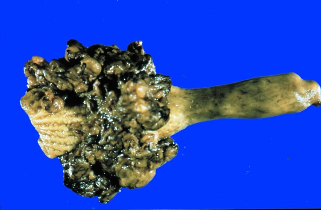
Macroscopic findings

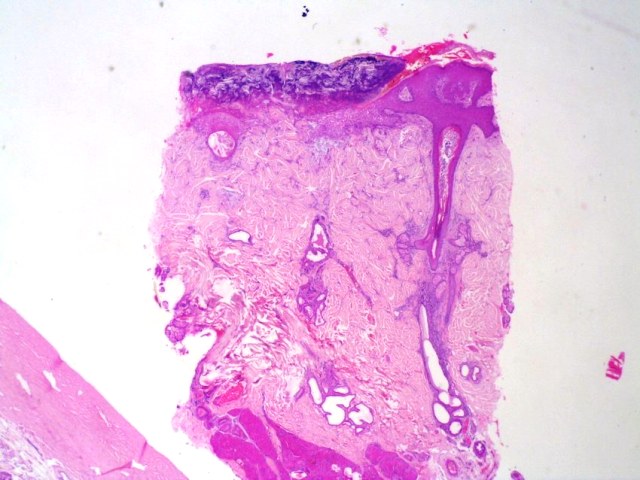
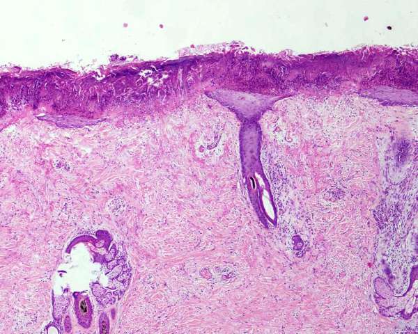
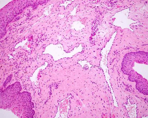
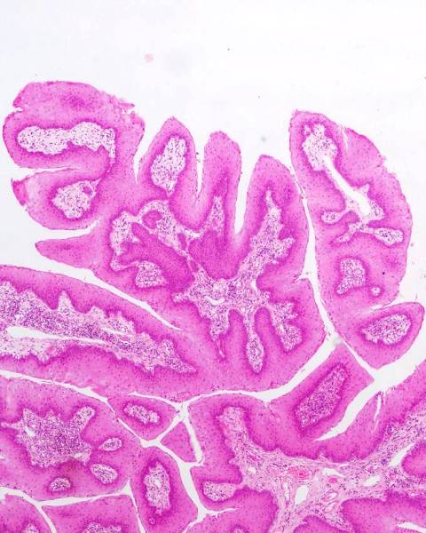
The lesions of transmissible venereal tumours are exophytic multinodular proliferations in the preputial cavity, often attached to the junction between the inner sheath of the prepuce and penile epithelium. The size can vary considerably from small nodules to large fungating masses that cause preputial swellings.

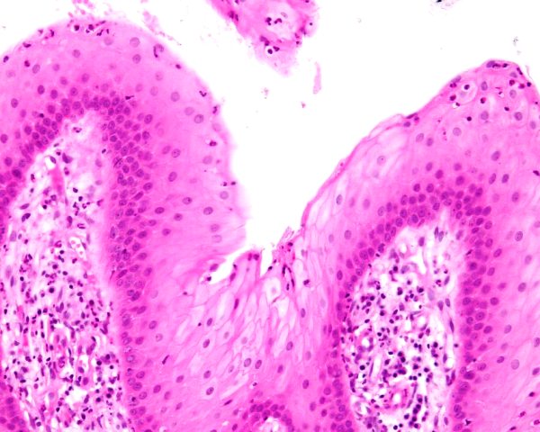
Figure : Transmissible venereal tumour, penis and prepuce

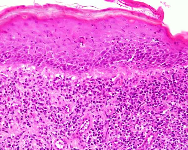
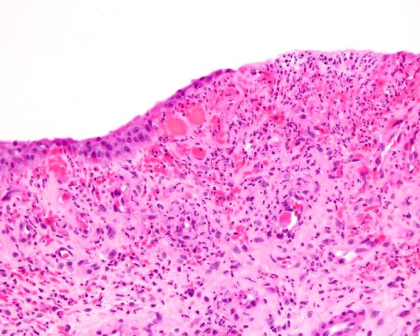
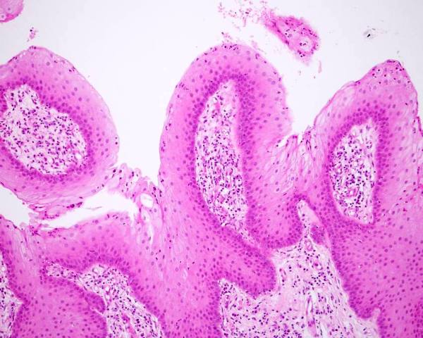
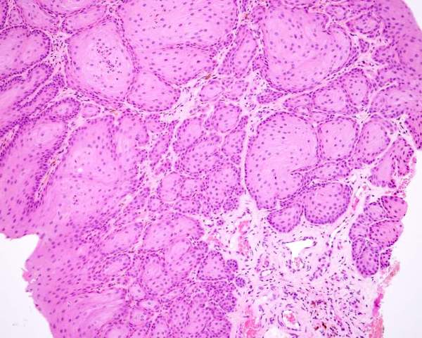
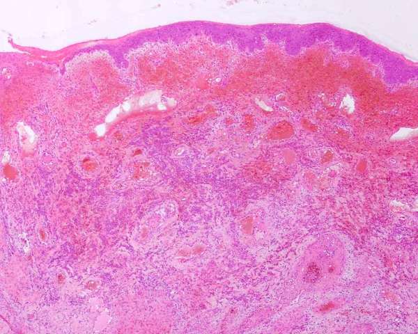
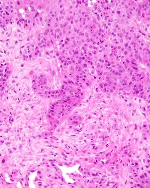
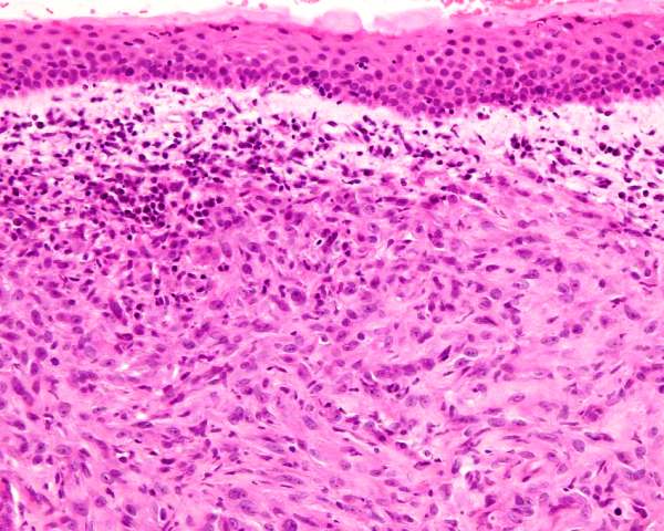
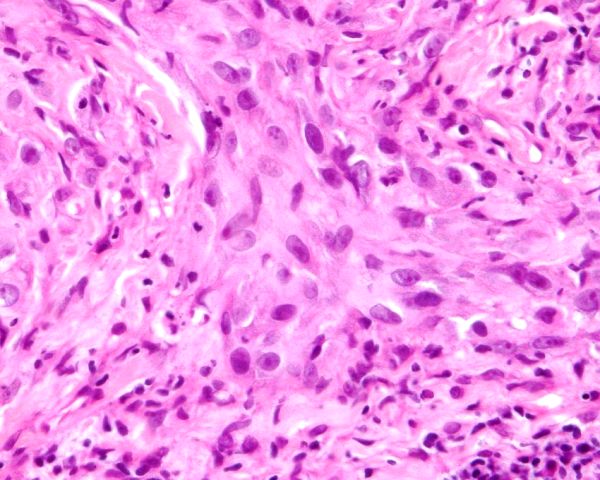
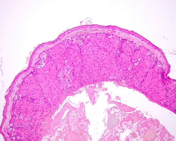
Microscopic findings

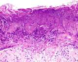
Transmissible venereal tumours are round cell tumours. They are locally infiltrative, and the cells are often arranged in packets with a fine stroma. The cells have well defined cytoplasmic boundaries, abundant often clear cytoplasm and a central round to indented nucleus. Mitoses are numerous. Some have fine vacuoles around the cytoplasmic membrane , and this feature is particulary prominent in cytological specimens. Cells such as macrophages and lymphocytes may be found in the neoplasms.

The differential diagnoses include lymphoma, histiocytic tumour, mast cell tumour and plasmacytoma.

Immunohistochemistry

Sandusky et al (1997) stained 4 CTVT with S-100, kappa and lambda light chains, alpha-1-antitrypsin, alpha-1-antichymotrypsin, LCA, neuron-specific enolase, keratin, cytokeratin, muramidase, and vimentin. The CTVT were consistently vimentin positive

Mozos et al (1996) reported on 25 CTVT stained for human lysozyme, human alpha-1-antitrypsin, CD3, vimentin, human keratins, human lambda light chains, canine immunoglobulins IgG, IgM, and S-100. Lysozyme was immunoreactive in 10/25 alpha-1-antitrypsin in 14/25, and vimentin in 25/25 CTVT. All were negative to keratins, S-100, lambda light-chain, IgG, IgM, and CD3.

Marchal et al (1997) reported CTVT stained for "lysozyme, ACM1 antigen, vimentin, neuron-specific enolase, glial fibrillary acidic protein, desmin, alpha smooth muscle actin, CD3, IgG, kappa and lambda light chains, and keratin. Lysozyme immunoreactivity was detected in all cases, ACM1 antigen in 11 of 14, neuron-specific enolase in 11 of 14, vimentin in 10 of 14, glial fibrillary acidic protein in 4 of 14 and desmin in 1 of 14. All the sections were negative to keratins, alpha smooth muscle actin and CD3, whereas in five cases, perivascular tumour cells contained Ig G, kappa and lambda light chains".

In summary then, CTVT are consistently vimentin positive, cytokeratin and S100 negative round cell tumours that do not stain for specific T or B lymphocyte markers and which stain for histiocytic markers.

Bright RM. Gorman NT, Probst CW, Goring CW. (1983) Transmissible venereal tumor of the soft palate in a dog. J Amer Vet Med Assoc 183: 893-894.

Cohen D (1973) The biological behaviour of the transmissible venereal tumour in immunosuppressed dogs. Europ J Cancer 9: 253-258

Cohen D (1978) The transmissible venereal tumor of the dog - a naturally occurring allograft? a review. Israel J Med Sc 14: 14-19.

Dass LL, Sahay PN (1989) Surgical treatment of canine transmissible venereal tumour - a retrospective study. Indian Vet J 66: 255-258.

Ganguly B, Das U, Das AK. Canine transmissible venereal tumour: a review. Vet Comp Oncol. 2016; 14: 1-12

Higgins DA (1966) Observations on the canine transmissible Venereal Tumour as seen in the Bahamas. Vet Rec 79: 67-71.

Hsiao YW, Liao KW, Hung SW, Chu RM (2002) Effect of tumor infiltrating lymphocytes on the expression of MHC molecules in canine transmissible venereal tumor cells. Vet Immunol Immunopathol. 87(1-2):19-27.

R. M. Kabuusu, D. F. Stroup, C. Fernandez (2010) Risk factors and characteristics of canine transmissible venereal tumours in Grenada, West Indies. Vet Comp Oncology 8: 50-55.

Liao KW, Hung SW, Hsiao YW, Bennett M, Chu RM (2003) Canine transmissible venereal tumor cell depletion of B lymphocytes: molecule(s) specifically toxic for B cells. Vet Immunol Immunopathol 92(3-4):149-162.

Marchal T, Chabanne L, Kaplanski C, Rigal D, Magnol JP.(1997) Immunophenotype of the canine transmissible venereal tumour. Vet Immunol Immunopathol. 57(1-2):1-11.

Marino G, Gaglio G, Zanghì A. (2012) Clinicopathological study of canine transmissible venereal tumour in leishmaniotic dogs. J Sm An Pract 2012,

Mello-Martins MI, Ferreira de Souza F, Gobello C (2005) Canine Transmissible Venereal tumor: etiology, pathology, diagnosis and treatment. In: Recent Advances in Small Animal Reproduction. Concannon PW, England G, Verstgegen J, Linde-Forsberg (Eds) www.ivis.org.

Milo J, Snead E. A case of ocular canine transmissible venereal tumor.Can Vet J. 2014; 55: 1245-1249.

Mozos E, Mendez A, Gomez-Villamandos JC, Martin De Las Mulas J, Perez J. (1996) Immunohistochemical characterization of canine transmissible venereal tumor. Vet Pathol33(3):257-263

Mukaratirwa S, Gruys E. (2003) Canine transmissible venereal tumour: cytogenetic origin, immunophenotype, and immunobiology. A review. Vet Q. 25(3):101-111.

Murchison EP, Wedge DC, Alexandrov LB, Fu B, Martincorena I, Ning Z, Tubio JM, Werner EI, Allen J, De Nardi AB, Donelan EM, Marino G, Fassati A, Campbell PJ, Yang F, Burt A, Weiss RA, Stratton MR. Transmissible [corrected] dog cancer genome reveals the origin and history of an ancient cell lineage. Science. 2014; 24: 343, 437-440.

Murgia C, Pritchard JK, Kim SY, Fassati A, Weiss RA. (2006) Clonal origin and evolution of a transmissible cancer Cell. 126(3):477-487.

Nak D, Nak Y, Cangul IT, Tuna B. (2005) A clinicopathological study on the effect of vincristine on transmissible venereal tumour in dogs. J Vet Med 52: 366-370.

Paia Chien-Chun, Kuob Tzong-Fu, Maoc Simon J.T. , Chuanga Tien-Fu, Lina Chen-Si, Chua Rea-Min (2011) Immunopathogenic behaviors of canine transmissible venereal tumor in dogs following an immunotherapy using dendritic/tumor cell hybrid Veterinary Immunology and Immunopathology 2011 139: 187–199

Parent R, Teuscher E, Morin M, Buyschaert A. (1983). Presence of the canine transmissible venereal tumor in the nasal cavities of dogs in the area of Dakar (Senegal). Canadian Vet J 24: 287-288.

Rogers KS, Walker MA, Dillon HB (1998) Transmissible venereal tumor: a retrospective study of 29 cases. J Amer Anim Hosp Assoc 34: 463-470.

Sandusky GE, Carlton WW, Wightman KA. (1987) Diagnostic immunohistochemistry of canine round cell tumors. Vet Pathol. 24(6):495-499

Stockmann D, Ferrari HF, Andrade AL, Cardoso TC, Luvizotto MCR. (2011) Detection of the tumour suppressor gene TP53 and expression of p53, Bcl-2 and p63 proteins in canine transmissible venereal tumour. Vet Comp Oncol 2011, 9: 251-259

Strakova A, Baez-Ortega A, Wang J, Murchison EP. Sex disparity in oronasal presentations of canine transmissible venereal tumour. Vet Rec. 2022 Jul 3:e1794.

Stockmann D, Ferrari HF, Andrade AL, Cardoso TC, Luvizotto MCR. (2011) Detection of the tumour suppressor gene
TP53 and expression of p53, Bcl-2 and p63 proteins in canine transmissible venereal tumour. Vet Comp Oncol 2011, 9: 251-259

Thornburn MJ, Gwynn RVR, Ragbeer MS, Lee BI. (1968) Pathological and cytogenetic observations on the naturally occurring canine venereal tumour in Jamaica (Sticker's tumour). British J Canccer 22: 720-727.

Weber WT, Nowell PC, Hare WCD (1965) Chromosome studies of a transplanted and primary canine venereal sarcoma. J Nat Cancer Inst 35: 537-547.

Wright DH, Peel S, Cooper EH, Hughes DT. (1970) Transmissible venereal sarcoma of dogs. A histochemical and chromosomal analysis of tumours in Uganda. Rev Europ etudes Clin Et Biol 15: 155-160.

Chondrosarcoma

Davis and Holt (2003) reported a case of two chondrosarcomas of the urethra

Davis GJ, Holt D. (2003) Two chondrosarcomas in the urethra of a German shepherd dog. J Small Anim Pract. 44(4):169-171


Haemangiosarcoma

There is one case of a penile haemangiosarcoma in the YagerBest Histovet Database


Lipoma

A single case of lipoma of the urethra was found in a dog with urethral obstruction in the penile urethra. The lesion was only microscopic, and was a nodule of well differentiated adipose forming an expansile lesion that compressed the penile urethra.

Lymphoma

Lymphoma of the penis and prepuce is a very rare manifestation of lymphoma. Lymphoma is typically a multisystemic disease, but there are several reports of the penis being involved primarily or as the first manifestation. Struble et al (1997) reported a case where there was obstruction of the penile urethra by a T cell lymphoma (Struble et al 2001). The case reported by Michels et al (2001) also had urethral obstruction, but there was also paraphimosis. Confirmation by immunohistochemistry or cytogenetics should be done.

It is critical that differentiation from Canine Transmissible Venereal Tumour be done by immunohistochemistry and cytogenetics as many pathologists mistake CTVT for lymphoma in non endemic areas.

Michels GM, Knapp DW, David M, Lantz GC, Munjar TA, Blevins WE, DeNicola DB.(2001) Penile prolapse and urethral obstruction secondary to lymphosarcoma of the penis in a dog. J Am Anim Hosp Assoc.37(5):474-477

Struble AL, Lawson GW, Ling GV. (1997) Urethral obstruction in a dog: an unusual presentation of T-cell lymphoma. J Am Anim Hosp Assoc. 33(5):423-426.

Ossifying fibroma of the os penis

Mirkovic et al (2004) reported a dog with an ossifying fibroma of the os penis. The proximity of the os penis to the urethra resulted in urinary obstruction.

Mirkovic TK, Shmon CL, Allen AL (2004) Urinary obstruction secondary to an ossifying fibroma of the os penis in a dog. J Am Anim Hosp Assoc. 40(2):152-156.

Osteosarcoma of os penis

Bleier et al (2003) reports on a case of osteosarcoma of the os penis in an older dog with urinary difficulties and blood in the urine. A mass developed in the inguinal region 2 months after surgery, but no further diagnostics were done.

Peppler et al (2009) reports on a dog with osteosarcoma of the os penis. Metastases were found 12 months after initial surgery.

Webb et al (2009) reports on a dog with a multilobular osteosarcoma (osteochondrosarcoma) in the os penis of a dog with dysuria. the neoplasm reoccurred twice.


Bleier T, Lewitschek HP, Reinacher M. (2003) Canine osteosarcoma of the penile bone. J Vet Med A 50: 397-398.

Peppler C, Weissert D, Kappe E, Klump S, Kramer M, Reinacher M, Neiger R (2009) Osteosarcoma of the penile bone (os penis) in a dog. Australian Vet J 87: 52-55

Webb JA, Liptak JM, Hewitt SA, Vince AR (2009) Multilobular osteochondrosarcoma of the os penis in a dog. Canadian Vet J 50: 81-84

Mesenchymoma

Root Kustritz and Fick (2007) in their Theriogenology Question of the Month report on a neutered dog that developed a 'benign mesenchymoma' of the os penis in a dog.

Root Kustritz MV and Fick JL (2007) Theriogenology Question of the Month. J Amer Vet Med Assoc 230: 197-198

Plasmacytoma

There is one plasmacytoma of the penis recorded in the YagerBest Histovet database.

Soft tissue sarcoma

Two cases of invasive soft tissue sarcoma are in the Yager-Best Histovet database. Each was a high grade lesion composed of infiltrative spindle shaped cells.

Figure : Penile soft tissue sarcoma.

Figure : Penile soft tissue sarcoma


Congenital anomalies

Hypoplasia and hypertrophy

A congentially small penis (also called an infantile penis) is occasionally seen (Ortega-Pacheco et al 2006). Micropenis or hypoplasia of the penis is a small penis. Megalopenis is an unusually large penis. Bloom (1954) reports both of these anomalies

Bloom F (1954) Pathology of the dog and cat - The genitourinary system, with clinical considerations. American Veterinary Publications, Inc, Evanston Illinois. p265

Ortega-Pacheco A, Rodríguez-Buenfil JC, Segura-Correa1 JC, Bolio-Gonzalez ME, Jiménez-Coello M and Linde Forsberg C. (2006) Pathological Conditions of the Reproductive Organs of Male Stray Dogs in the Tropics: Prevalence, Risk Factors, Morphological Findings and Testosterone Concentrations. Reprod Dom Anim 41 :429-437

Meilan J. (2006) Rare preputial anomaly and its surgical correction in a dog. Vet Rec.158(19):672.

Hypospadias and fistulas

Hypospadias is when the urethra opens on the ventral surface of the penis (or on the perineum). It is the result of a failure of complete closure of the urethra during development.

This is one of the more common anomalies of the reproductive tract of dogs. Ader and Hobson (1978) reported 3 cases in dogs. Hayes and Wilson (1986) reviewed medial records of 17 North American veterinary schools and identified 66 cases of dogs with hypospadias. 59 were phenotypic males. Boston terriers were overrepresented. Jurka et al (2009) reported 6 cases, one of which was a hermaphrodite. Two were perineal, 2 were penile and one was so called glandular in type. The hermaphrodite was scrotal in type.Adelsberger and Smeak (2009) report a single case in a Boston terrier that they repaired surgically.

There are also many different fistulas where the urethra opens into other structures - these include urethrorectal and urethroperineal fistulas. Some of these do not involve the penis.

Adelsberger ME, Smeak DD (2009) Repair of extensive perineal hypospadias in a Boston terrier using tubularized incised plate urethroplasty. Canadian Vet J. 50: 937-942

Ader PL, Hobson HP (1978) Hypospadias: a review of the veterinary literature and a report of three cases in the dog. J Amer Animal Hosp Assoc 14: 721-727. as quoted by McEntee (1990)

Hayes HM, Wilson GP (1986) Hospital incidence of hypospadias in dogs in North America. Vet Rec 118: 605-606.

Lefebvre R, Lussier B (2005) A clinical case of hypospadias in a dog. Canadian Vet J 46: 1022-1025.

Jurka P, Galanty M, Zielinska P, Max A, Sysa P. (2009) Hypospadias in six dogs Vet Rec 164: 331-333

Balanopreputial fold or band (Persistent frenulum)

The internal sheath of the prepuce and the epithelium of the penis are fused in neonates, and separate after birth. In some animals this only occurs at puberty. Failure of a portion to separate results in a balanopreputial band. A band from the frenulum of the penis to the prepuce is a persistent frenulum. The frenulum is located on the ventral surface of the free portion of the penis, but not the head of the penis. According to the NAV, the part of the penis of the dog within the prepuce is the head of the penis (including the bulb) so a true frenulum would be difficult! There are many individual reports of a 'persistent frenulum' in the literature, and the recognition and treatment is simple and does not require the expertise of a surgical pathologist.

There is a report of retained bands, called balanopreputial folds or bands that caused phimosis and accumulation of fluid in the cavity of the prepuce (Olsen and Salwei 2001)


Balke J. (1981) Persistent penile frenulum in a cocker spaniel. Vet Med Small Anim Clin. 76(7):988-990

Begg TB (1963) Persistent penile frenulum in the dog. Vet Rec 75: 930-931

Belkin PB (1969) persistence of penile frenulum in a dog Mod Vet Pract 50(11): 80

Barrand KR (1999) What is your diagnosis? Persistent penile frenulum. J Small Anim Pract. 40(3):105, 142

Hutchison JA. (1973) Persistence of the penile frenulum in dogs. Can Vet J. 14(3):71

Joshua AO (1962) Persistence of the penile frenulum in a dog. Vet Rec 74: 1550-1551

Olsen D, Salwei R.J Surgical correction of a congenital preputial and penile deformity in a dog. J Am Anim Hosp Assoc 37(2):187-192

Ryer KA. (1979) Persistent penile frenulum in a Cocker Spaniel Vet Med Small Anim Clin. 74(5):688

Miscellaneous conditions

Absence of preputial oriface

Elam and Randle (1952) report a condition where the preputial orifice was missing in a 5 week old puppy.

Chaffee VW, Knecht CD. (1975) Canine paraphimosis: sequel to inefficient preputial muscles. Vet Med Small Anim Clin. 70(12):1418-1420.

Elam CW, Randle PO (1952) Peculiar preputial condition in a five week old puppy. Vet Rec 64: 98.

Curved os penis

Johnston (1965) reports a dog with a curved os penis

Johnston DE (1965) Repairing lesions of the canine penis and prepuce. Mod Vet Pract 46: 39-46.

Diphalia

Diphalia is a double penis. This is reported by Johnston et al (1989).

Johnston SD, Bailie NC, Hayden DW, Johnston GR, Osborne CA. (1989) Diphallia in a mixed-breed dog with multiple anomalies. Theriogenology. 31(6):1253-1260.

Membranous urethra

Kipnis (1974) reports an unusual of the penile urethra where the urethra was membranous.

Kipnis RM (1974) Membranous penile urethra and preputial abnormality in a dog. Vet Med Small Anim Pract 69: 750-751

Urethral duplication

Tobias and Barbee (1995) reported on a dog that had multiple anomalies that included a urethral duplication.

Duffey et al (1998) reports on a dog that had intermittent incontinence and was found to have an incomplete urethral duplication of the penile urethra that had an accompanying cyst.

Tobias KS, Barbee D (1995) Abnormal micturition and recurrent cystitis associated with multiple congenital anomalies of the urinary tract in a dog. J Am Vet Med Assoc 207(2):191-193

Duffey MH, Barnhart MD, Barthez PY, Smeak DD (1998) Incomplete urethral duplication with cyst formation in a dog. J Amer Vet Med Assoc 213: 1287-1288.

Disease of the external sheath of the prepuce

The external sheath of the penis is haired skin, and as such shares its diseases with the rest of the skin. This is the domain of the dermatologist and the dermatopathologist, and is beyond this review. There are some diseases that are unique to this area and these will be highlighted.

Mast cell tumour

There is a general belief that mast cell tumours of the scrotum and inguinal area are more aggressive and 'malignant' than those elsewhere on the body. Cahalane et al (2004) attempted to determine if mast cell tumors of the inguinal and perineal skin were prognostically different to skin elsewhere. There was no difference in their series.

Cahalane AK, Payne S, Barber LG, Duda LE, Henry CJ, Mauldin GE, Frimberger AE, Cotter SM, Moore AS. (2004). Prognostic factors for survival of dogs with inguinal and perineal mast cell tumors treated surgically with or without adjunctive treatment: 68 cases (1994-2002). J Amer Vet Med Assoc 225: 401-408.

Hepatoid gland tumours (preputial gland)

The prepuce of dogs may contain hepatoid glands. These glands are well known around the anus where they are called perianal glands. Neoplasms of perianal hepatoid glands are common and well recognized.

In the YagerBest Histovet database there are 12 cases where masses from the prepuce were identified as being of hepatoid origin. Three of these were invasive and met the criteria of being carcinomas and one of the 3 was a mixed tumouor with osteoid formation. These tumours had the typical appearance of hepatoid tumours.

)

Figure : Hepatoid adenoma of the prepuce

Figure : Hepatoid adenoma of the prepuce

Figure : Hepatoid adenoma of the prepuce

Selected references of lesions of the external prepuce

Galofaro V, Rapisarda G, Ferrara G, Iannelli N (2006) Glomangioma in the Prepuce of a Dog. Reprod Dom Anim 41 568