This page is part of the site called Surgical Pathology of the Canine Female Reproductive Tract by

Dr Rob Foster

OVC Pathobiology

University of Guelph

© All material is copyright and should not be used without the expressed permission of the author.


Disease of the Canine Uterus

Table of Contents


General considerations

Lesions of the uterus of dogs are common. The majority are found at ovariohysterectomy as 'incidental' lesions, or are found when there is a vaginal discharge. The submissions to a surgical biopsy service will not reflect the overall occurrence of diseases of the uterus, as most of the readily recognised lesions will not be submitted. Pyometra is a good example. Many of the submissions are for 'masses' in the uterus, such as neoplasia, adenomyosis, polyps or cystic structures. The approach to the lesions taken here reflects the occurrence in the Yager Best database.

The following table gives some indication of the occurrence of lesions and what to expect in submissions to a surgical pathology service.

Hyperplasia  

Cystic endometrial hyperplasia

135

spontaneous endometrial hyperplasia

36

Endometrial polyp

11

Adenomyosis

56
endometrial cyst
6
hyperoestrogenism

6


Neoplasia

leiomyoma
16
fibroleiomyoma
2

leiomyosarcoma

5
lymphoma
1
endometrial stromal tumour
1
Mucometra/hydrometra
30
   
Serosal inclusion cysts
17
   
Endometritis/pyometra
125
   
Uterine stump inflammation, hyperplasia
14
 
 
Anomalies

Segmental aplasia

4

Remnants and duplications

10
   
Hernia
1
Haemorrhage
5
Melanosis
1
   

General studies on Canine Uterine Disease


Maya -Pulgarin et al (2018) reported on histological findings in the uterus of 76 dogs with macroscopic lesions visible during elective ovariohysterectomy of a population of 3,600 dogs. They found  cystic endometrial hyperplasia, periglandular fibrosis, lymphoplasmocytary endometritis, and adenomyosis in 19.7%, 14.5%, 4.0%, and 2.6%, respectively.

Maya-Pulgarin D, Gonzalez-Dominguez MS, Aranzazu-Taborda D, Mendoza N, Maldonado-Estrada JG. Histopathologic findings in uteri and ovaries collected from clinically healthy dogs at elective ovariohysterectomy: a cross-sectional study. J Vet Sci. 2017; 18: 407-414.


Generalised endometrial atrophy

Dockweiller et al (2017) reported on a dog that had no evidence of progesterone influence (essentially atrophy) and no progesterone receptors (PR) were found by immunohistochemistry. The assumption was that infertility was due to PR absence.


Dockweiler JC, Cossic B, Donnelly CG, Gilbert RO, Buckles E, Cheong SH. Infertility associated with the absence of endometrial progesterone receptors in a bitch. Reprod Domest Anim. 2017; 52: 174-178.

Generalized endometrial hyperplasia

Cystic endometrial hyperplasia and the decidual reaction.

The majority of pathologists recognise cystic endometrial hyperplasia. Recent and not so recent research has shown that not all cystic endometrial hyperplasia are created equal - and there are subtypes. While the cynical will say 'what does it matter?' or 'So?', there are pathogenetic differences and variations in outcome.

Bitches that have estrous cycles but do not become pregnant may develop spontaneous cystic hyperplasia. These may subsequently develop endometritis and pyometra. Mechanical cystic endometrial hyperplasia occurs when there is foreign material within the uterus (usually bacteria) so endometrial hyperplaisa can occur because of infection. Dogs treated with progesterone develop cystic endometrial hyperplasia, and there is some hyperplasia in the early phase of treatment with oestrogen. Dogs with chronic pyometra may not have endometrial hyperplasia. Confused?

My approach is to try to separate out the various forms of cystic endometrial hyperplasia based on pathogenesis. At the same time it is important to recognise that the most common situation is that outlined by Dow (1957) where there are 4 stages recognised in what is commonly called the cystic endometrial hyperplasia-pyometra complex. Hopefully there will be some sense made to all of this in the next sections.

Spontaneous generalized cystic endometrial hyperplasia (CEH)

Cystic endometrial hyperplasia is one of the most well recognised conditions of the canine uterus. It is one of the least understood. For decades, this condition was recognised to have a hormonal basis, and to arise when bitches did not become pregnant during oestrus. The common association with endometritis and pyometra led to the combination of conditions to be called CEH-pyometra. Unravelling this association has been difficult as it is so entrenched.

Pathogenesis

Just why the endometrium undergoes hyperplasia is not known. An early association between endometrial hyperplasia and pyometra was made (Dow 1957), but for this section, we will concentrate on uncomplicated CEH. Dow (1957) surveyed 100 cases and classified the animals into 4 groups. He recognised that there was a group of 23 dogs that had what he termed uncomplicated cystic hyperplasia. These bitches had a mucoid discharge during metoestrus. These were therefore likely to have spontaneous cystic endometrial hyperplasia.

Normal cyclic changes in the endometrium are under the control of ovarian steroidal hormones including oestrogen and progesterone. Without these hormones, the endometrium atrophies. The dog is unique in having either one or two oestrus periods per year, and they have a prolonged dioestrus and anoestrus. The dioestral period is when the corpus luteum is present (also called the luteal phase), so the uterus has a long period of exposure to progesterone.

The cyclical changes in bitches result in changes in the endometrium but a surprising lack of cell death or proliferation in the epithelium as De Bosschere et al (2002) found when they stained for apoptosis and the proliferation marker Ki67. When dogs are not bred, have infertile matings, or are treated with progestagens for prolonged periods (Kim and Kim 2005), cystic endometrial hyperplasia may develop. Progestagen is required to maintain the endometrium, and withdrawl of progestagen, even in the face of uterine irritation or hyperplasia, will result in its dissappearance (Chen et al 2006).

Cystic endometrial hyperplasia does not appear to be influenced by growth hormone produced in the mammary gland when progesterone is administered (Bhatti et al 2006).

In a study of endometrial hyperplasia and the influence of treatment with progesterone, Von Berky and Townsend (2003) found that of 42 bitches treated with medroxyprogesterone acetate, 45% developed CEH whereas only 5% of 133 untreated had lesions.

Estrogen and progesterone receptor expression does not appear to be involed in the pathogenesis (De Bosschere et al 2002)

Macroscopic appearance

A uterus with cystic endometrial hyperplasia is thicker and there developed an exagerated nodularity. The area of the endometrium compared to the area of the myometrium increases.

Microscopic appearance

Histological changes in the endometrium with hyperplasia varies from mild to severe. Mild hyperplasia is a thickening of the endometrium with elongation of the glands. As this becomes more greater, the glands become disorganised and rather than straight, become convoluted so that the cross sections vary. The stroma becomes more prominent. There is dilation of the glands with the formation of variably sized cysts. It is the variability of the size of cystic dilation of glands that is characteristic.

In general, uncomplicated spontaneous endometrial hyperplasia does not have inflammatory cell infiltrates, nor is the surface epithelium vacuolated or hypertrophied - this occurs with there is a luminal content like mucin, pus, or other exudates.

Bhatti SF, Rao NA, Okkens AC, Mol JA, Duchateau L, Ducatelle R, van den Ingh TS, Tshamala M, Van Ham LM, Coryn M, Rijnberk A, Kooistra HS. (2006) Role of progestin-induced mammary-derived growth hormone in the pathogenesis of cystic endometrial hyperplasia in the bitch. Domest Anim Endocrinol (in press)

Chen YM, Lee CS, Wright PJ. (2006) The roles of progestagen and uterine irritant in the maintenance of cystic endometrial hyperplasia in the canine uterus. Theriogenology. 66(6-7): 1537-1544.

De Bosschere H, Ducatelle R, Tshamala M. (2002) Is mechanically induced cystic endometrial hyperplasia (CEH) a suitable model for study of spontaneously occurring CEH in the uterus of the bitch? Reprod Domest Anim 37(3): 152-157.

De Bosschere H, Ducatelle R, Vermeirsch H, Simoens P, Coryn M. (2002) Estrogen-alpha and progesterone receptor expression in cystic endometrial hyperplasia and pyometra in the bitch. Anim Reprod Sci 70: 251-259

Dow C. (1957) The cystic hyperplasia-pyometra complex in the bitch. Vet Rec 69: 1409-1414.

Kim KS, Kim O. (2005) Cystic endometrial hyperplasia and endometritis in a dog following prolonged treatment of medroxyprogesterone acetate. J Vet Sci. 6(1): 81-82.

Von Berky AG, Townsend WL. (1993) The relationship between the prevalence of uterine lesions and the use of medroxyprogesteroe acetate for canine population control. Aust Vet J 70: 249-250.


Noakes DE, Dhaliwal GK, England GCW. (2001) Cystic endometrial hyperplasia/pyometra in dogs: a review of the causes and pathogenesis. J. Reprod Fert Suppl 57: 395-406 2001 (review)


Foreign body (mechanical) associated endometrial hyperplasia

Endometrial hyperplasia is induced by foreign materials such as a silk suture, olive oil, barium, uterine grafts or other materials. There is proliferation of the endometrium with a dramatic increase in the proliferation marker Ki67 (De Bosschere et al 2002). This proliferation appears to be identical to that which occurs when the endometrium comes in contact with bacteria (in pyometra) or fertilized ova (in early pregnancy) in both its appearance and in progesterone and oestrogen receptor expression. (De Bosschere et al 2002). It is different in receptor expression to naturally occurring cystic endometrial hyperplasia. Progesterone and oestrogen receptor expression is reduced in the epithelial cells in the mechanically stimulated condition, whereas it is increased in the naturally occurring disease. Progesterone receptors are increased and oestrogen receptors are decreased in the stroma of mechanically induced hyperplasia whereas both are increased in the stroma in cases of the naturally occuring disease.

Kainz (1914) was one of the first to recognize that endometrial hyperplasia occurs at the site of traumatic stimulation, in his case, a suture line. He recognised the reaction as being similar to the normal placental site.

Hadley JC (1975) was the first to introduce the possibility of mechanical involvement in the development of cystic endometrial hyperplasia. Hadley (1975) reported that bitches subjected to serial uterine biopsies developed cystic endometrial hyperplasia and endometritis, whereas those that had uterine palpation alone did not.

Koichi Nomura, from the University of Osaka Prefecture, performed research into endometrial hyperplasia by firstly inducing pyometra by inoculating uteri after estrus with E. coli from a dog that had pyometra. He observed that infection with E. coli induced endometrial hyperplasia and suspected that hyperplastic changes occur in the uterus with any stimulus (Nomura 1983). Endometrial hyperplasia was subsequently induced by intrauterine scratching (Nomura et al 1990). The histological finding was that of dilation of the endometrial glands (particularly of the basal region of the glands) and hyperplasia to give a 'swiss cheese endometrium'. This change was different to that reported as associated with long term administration of progesterone by Dow (1959), where the hyperplasia and dilation began at the tubular portion of the endometrial gland. Nomura (1994) continued by attempting to produce hyperplasia (or deciduoma) with noninfectous materials in bitches in metoestrus. These included scratching the endometrium with a stainless steel wire and by placing a transmural silk suture. Insertion of olive oil in the lumen produced a mild hyperplasia and insertion of saline had no effect. A marked decidual reaction was seen with the silk suture. This included irregular but marked dilation of the glands, and papillary hyperplasia of the luminal epithelium (Nomura 1995a). The changes induced by olive oil were similar but less marked. There was hyperplasia of the superficial glands, and dilation of the basal portion of the glands. The surface had papillary hyperplasia.


De Bosschere H, Ducatelle R, Tshamala M. (2002) Is mechanically induced cystic endometrial hyperplasia (CEH) a suitable model for study of spontaneously occurring CEH in the uterus of the bitch? Reprod Domest Anim 37(3): 152-157.

Dow C (1959) Experimental reproduction of cystic hyperplasia -pyometra complex in the bitch. J Pathol Bacteriol 78: 267-278.

Kainz K (1914) Arch Mikr Anat 84: 122-141. As quoted by

Nomura K (1983) Canine pyometra with cystic endometrial hyperplasia experimentally induced by E. coli inoculation. Jpn J Vet Sci 45: 237-240.

Nomura K (1994) Induction of a deciduoma in the dog. J Vet Med Sci 56: 365-369

Nomura K (1995a) Histological evaluatoin of canine deciduoma induced by silk suture. J Vet Med Sci 57: 9-16.

Nomura K (1995b) Histological evaluation of the canine decidual reactions induced by intraluminal injection of olive oil. J Vet Med Sci 57: 71-74

Nomura K, Kawasoe K and Shimada Y (1990). Histological observation of canine cystic endometrial hyperplasia induced by intrauterine scratching. Jpn J Vet Sci 52: 979-983.

Progesterone induced endometrial hyperplasia

Progesterone treatment of ovarectomised dogs

Progesterone administration to bitches produces endometrial changes. Dow (1959) gave ovarectomised bitches progesterone alone and found that in the first 10 days there were changes whereby the superficial epithelium became a layer of tall columnar cells with a vacuolated cytoplasm and large vesicular nuclei. The endometrial glands, particularly the tubular portion, were lined by similar tall vacuolated cells. As time and treatment continued, the changes became more exagerated so that after 40 days, there is a marked increase in the number of glands and there were polypoid projections into the lumen. The basal portion of the glands were less affected. By 60 days, the all endometrial glands and all parts (basal and tubular) were dilated, but to varying degrees. The larger the cystic dilation, the thinner the epithelial cells lining that portion. Those of the luminal epithelium were tall columnar and highly vacuolated. The stroma did not appear to he increased in amount. The changes developed quicker in bitches that were pretreated with oestrogen.

Dow (1959) then examined the uteri of bitches that were given cycles of oestrogen followed by progesterone. After one cycle, there was little change. After 2 cycles, there was some dilation of glands, but no cystic change. After 4 cycles, all 4 dogs developed pyometra and had cystic dilation of glands. Dogs given 4 cycles of treatment and developed endometritis had treatment withdrawn and the uteri returned to normal.

Progesterone treatment of intact dogs

Withers and Whitney (1967) reported on a large number of dogs given medroxyprogesterone acetate (MPA). It was used to suppress oestrus, which it did in 83 of 86 bitches. 1 of the 86 had endometritis and 2 developed cystic endometrial hyperplasia. Three bitches with pyometra were treated also and all 3 died.

Von Berky and Townsend (1993) examined 42 uteri of dogs given MPA for estrus control. These were compared to 133 other doge in the same population. 45% of MPA treated bitches had uterine lesions whereas 5% of the untreated dogs had lesions. The lesions were endometrial hyperplasia and pyometra, but no other details.


Von Berky AG, Townsend WL (1993) The relationship between the prevalence of uterine lesions and the use of medroxyprogesterone acetate for canine population control. Aust Vet J 70: 249-250.

Withers AR, Whitney JC (1967) The response of the bitch to treatment with medroxyprogesterone acetate. J Small Anim Pract 8: 265-271

Estrogen induced endometrial hyperplasia

Ovarectomised bitches

Attempts to produce endometrial hyperplasia by administration of estrogen alone have been both successfull and unsuccessful. Dow (1959) failed to induce endometrial hyperplasia in ovarectomised bitches. He reviewed the early literature and reported on one author who reported hyperplasia from hyperestrogenism and 2 who failed to induce hyperplasia.

Dogs exposed and affected by estrogen replacement sprays or creams may develop hyperestrogenism and stump pyometra.


Sterman AA, Mankin KT, Barton CL. Stump Pyometra Secondary to Human Topical Estrogen Hormone Exposure in a Spayed Female Chihuahua. J Am Anim Hosp Assoc. 2019; 55: e55604


Intact bitches

When estrogen is given to bitches who are intact (have ovaries), estrogen administration induces endometrial hyperplasia. Jabara (1962) reported that the endometrium of stilboestrol treated dogs had the same appearance as bitches in oestrus and metoestrus, but after prolonged administration, the endometrium was 'degenerate' - it had marked glandular atrophy. Some developed endometritis.

Schwartz et al (1969) dosed 6 month old dogs with quinestrol for one year. They found that the dogs developed vaginal exudate, swollen mammary glands, enlarged nipples, vulval swelling and alopecia, especially at higher doses. At higher doses there was uterine enlargement with endometrial hyperplasia and myometrial hypertrophy.

Intact bitches with clinical hyperoestrogenism

Fiorito (1992) reported on a dog that had clinical signs of hyperoestrogenism including alopecia, hyperpigmentation and lichenification of the inguinal skin. The dog had multiple follicular cysts of both ovaries.

Fayrer-Hosken et al (1992) reports on a dog with clinical hyperoestrogenism that has follicular cysts of one ovary and cystic endometrial hyperplasia and pyometra.


Dow C (1959) Experimental reproduction of cystic hyperplasia -pyometra complex in the bitch. J Pathol Bacteriol 78: 267-278.

Fayrer-Hosken RA, Durham DH, Allen S, Miller-Liebl DM, Caudle AB. (1992) Follicular cystic ovaries and cystic endometrial hyperplasia in a bitch.
J Am Vet Med Assoc 201(1): 107-108.

Fiorito DA (1992) Hyperestrogenism in bitches. Compend Cont Edu 14: 727-729

Jabara AG (1962). Some tissue changes in the dog following stilboestrol administration. Aust J Exp Phil 40: 293-308.

Schwartz E, Tornaben JA, Boxill GC (1969) Effects of chronic oral administration of a long acting estrogen Quinestrol to dogs. Toxicol Applied Pharm. 14: 487-494.


Localized endometrial hyperplasia

Focal endometrial hyperplasia of pseudopregnancy (aka pseudo-placentation endometrial hyperplasia)

These are focal swellings of the uterus and they resemble placental sites, but without the presence of a conceptus. It is common for this to be misdiagnosed by pathologists as pregnancy, and the client goes in search of the invisible male that 'caused this outrage'. The swellings can be solitary or multiple.

The term deciduoma is increasingly being used in conjunction with swellings of the endometrium in dogs. A deciduoma is a swelling of decidual tissue, the tissue that 'falls off' - it is the endometrial tissue that forms during pregnancy or, in primates, during menstruation.

For this focal endometrial hyperplasia to occur, the dogs must be in dioestrus and have corpora lutea and progesterone in circulation. Hadley (1975) found that the serum oestrogen and progesterone concentration was within normal limits in single samples collected from dogs with false pregnancy and in dogs with pyometra.

It can be seen from the section on mechanically induced endometrial hyperplasia that the canine uterus is particularly adept at undergoing hyperplasia in response to luminal stimulation. It is not surprising that the uterus can attempt to form a placental site even though there is not conceptus. It is assumed that focal hyperplastic lesions that resemble placental sites are of this type. They may occur in pseudopregnancy. Pseudopregnancy is a physiological event that may be exagerated and clinically visible. Excellent reviews are available (Gobello et al 2001a, b). The pathology of the uterus in pseudopregnancy is documented by Whitney (1967). Clinically pseudopregnant dogs had diffuse thickening of the uterus and additionally, macroscopically visible swellings that corresponded to 2cm diameter regions of pronounced endometrial hyperplasia. The lumen of the affected uteri had a clear or cloudy blood stained fluid (see mucometra). Microscopically, the placenta like zones had long branching villous structures and the epithelial cells of the the lumen and the glands were large and foamy. The endometrial glands are dilated and contain secretion. Whitney (1967) reported that 3 of 6 dogs had inflammatory cells including neutrophils in these placental sites.

Arrighi et al (2007) reports on finding granulated lymphocytes in the interstitium of a dog with deciduoma. These cells have bilobed or reniform nuclei and had eosinophilic granules in their cytoplasm (and thus resembled eosinophils). The granules were PAS postive, and some were CD3+. They were CD74+ whereas eosinophils should be negative.

Schaffer and Gifford (2008) provides an excellent review of cystic endometrial hyperplasia, pseudo-placentation endometrial hyperplasia and other cystic conditions of the canine and feline uterus. They called this condition pseudo-placentation endometrial hyperplasia. This term is now gaining wider acceptance.

Sato (2011) published a case report that details a single case of pseudo-placentation endometrial hyperplasia.

Some papers confuse this condition with the hyperplasia of the endometrium that occurs with foreign body induced hyperplasia in a diestrus dog (Santana et al 2020).


Photomicrograph of localised endometrial hyperplasia of pseudopregnancy (pseudo-placentation endometrial hyperplasia) in a bitch (YB162911)


Arrighi S; Cremonesi F; Bosi G; Groppetti D, Pecile A. (2007) Characterization of a Population of Unique Granular Lymphocytes in a Bitch Deciduoma, Using a Panel of Histo- and Immunohistochemical Markers Vet Pathol 44: 521-524.

Gobello C, Concannon PW, Verstegen J (2001) Canine pseudopregnancy: a review. In Recent Advances in Small Animal Reproduction, PW Concannon, G England, J Verstegen (Eds) International Veterinary Information Service (www.ivis.org) Ithaca NY USA.

Gobello C, de la Sota RL, Goya RG (2001) A review of canine pseudocyesis. Reprod Dom Anim 36: 283-288.

Hadley JC (1975) Unconjugated oestrogen and progesterone concentrations in the blood of bitches with false pregnancy and pyometra. Vet Rec 96: 545-547

Sato Y. (2011) Pseudo-placentational endometrial hyperplasia in a dog. J Vet Diagn Invest 2011, 23: 1071-1074,

Santana CH, Santos DO, Trindade LM, Moreira LG, Paixão TA, Santos RL. Association of Pseudoplacentational Endometrial Hyperplasia and Pyometra in Dogs. J Comp Pathol. 2020; 180: 79-85.

Schlafer DH, A.T. Gifford AT. (2008) Cystic endometrial hyperplasia, pseudo-placentational endometrial hyperplasia, and other cystic conditions of the canine and feline uterus. Theriogenology 2008, 70: 349-358

Whitney JC (1967) The pathology of the canine genital tract in false pregnancy. J Small Anim Pract 8: 247-263

Adenomyomatous polyp

Adenomyomatous polyp is like an endometrial polyp in being a sessile often pedunculated mass in the uterus and arising from the endometrium. This differs from a regular endometrial polyp in having smooth muscle in the stroma. This stroma is mostly smooth muscle. These are very rare.

Nueangphuet P, Tanabe M, Izzati UZ, Fuke N, Hirai T, Yamaguchi R. Adenomyomatous uterine polyp in a Miniature Pinscher: histologic, immunohistochemical, and clinical aspects. J Vet Diagn Invest. 2022; 34: 102-106.

Endometrial polyps

Endometrial polyps are believed to develop from endometrial hyperplasia (Gelberg and McEntee 1984), as there is almost always some degree of endometrial hyperplasia in affected individuals. Polyps generally fall into 3 main categories - endometrial polpys (epithelium and stroma), endometrial stromal polyps (mostly stroma) and hypercellular polyps (so cellular you are tempted to call them neoplasia).

In the publication by Gelberg and McEntee (1984), polyps were found in 17 dogs. The largest was 20 cm diameter! Only 2 of these dogs did not have cystic endometrial hyperplasia. Gumber et al (2010) reported another large one and it was 17 x 9 x 4 cm and was hemorrhagic. The dog had concurrent pyometra.

Marino et al 2013 provided details of 21 dogs with endometrial polyps, including some as large as 25cm diameter! Uterine torsion was found in one case.

Nueangphuet et al (2022) described what they called an adenomatous polyp. Their polyp looked just like all the other endometrial polyps.

There are 18 cases of endometrial polyps in the YB database.

Macroscopic findings

Although polyps can be microscopic in size, most are recognised as swellings of the uterus. They may be single or multiple and occur anywhere in the uterus (Bloom 1954). They should have a pedicle, but some have a broad base. Most are solitary and, when the uterus is opened, appear as a nodule on a stalk. Intussusception or prolapse of the polyp can occur.

Microscopic findings

Polyps are composed of endometrial tissue identical to surrounding endometrium. They usually are composed of an outer layer of epithelium and stroma within which are distended uterine glands. In all locations, the cells, be they epithelium or stroma, are well differentiated, and do not have dysplastic features. The microscopic findings are identical to those of cystic endometrial hyperplasia. There is usually concurrent cystic endometrial hyperplasia. Smooth muscle may be found in the interstitium of the polyp.

Bloom (1954)

Gelberg HB, McEntee K. Hyperplastic endometrial polyps in the dog and cat. Vet Pathol 1984; 21: 570-573.

Gumber S, Springer N, Wakamatsu N. Uterine endometrial polyp with severe hemorrhage and cystic endometrial
hyperplasia–pyometra complex in a dog. J Vet Diagn Invest 2010 22:455–458

Marino G, Barna A, Zangh Catone G. Endometrial Polyps in the Bitch: a Retrospective Study of 21 Cases. J Comp Pathol 2013; 149: 410-416

Nueangphuet P, Tanabe M, Izzati UZ, Fuke N, Hirai T, Yamaguchi R. Adenomyomatous uterine polyp in a Miniature Pinscher: histologic, immunohistochemical, and clinical aspects. J Vet Diagn Invest. 2022; 34: 102-106.

Polypoid adenomyomatosis

Zanghi et al (2007) reports on a single case of a dog with a granulosa cell tumour and pyometra. There were multiple sessile or pedunculated lesions of the endometrium that had epithelial and stromal hyperplasia, with the latter containing a large amount of smooth muscle. The rest of the endometrium was atrophic and had squamous metaplasia. The lesions were called adenomyomatosis after a similar disease in women. Since reading this report, I have seen quite a few of these - previously grouping them with the endometrial polyps.

Endometrial polypoid adenomyomatosis.

Zanghi A, Catone G, Marino G, Quartuccio M, Nicotina PA. (2007) Endometrial polypoid adenomyomatosis in a bitch with ovarian granulosa cell tumour and pyometra. J Comp Pathol 136(1): 83-86.

Endometritis and pyometra

Inflammation of the endometrium is endometritis. Pyometra is when the uterus is filled with pus. There is a continuum in the degree of inflammation from the very mildest changes of endometritis through to the most severe cases of pyometra. The vast majority of cases are initiated when the cervix is open at estrus but there is also some evidence to support of haematological localisation of bacteria in the uterus.

It is typical to categorise endometritis into acute, subacute and chronic stages. I do not do this. I describe endometritis according to the cell types present and the degree of fibrosis.

Endometrial biopsy is now being used more and more to identify changes in the endometrium. Highly experienced individuals have reported the histological findings. Endometrial biopsy in bitches requires skill in performing transcervical samples. So far, endometrial biopsy in dogs have only been reported in bitches with fertility problems. This creates problems with suitable controls.

Gogny et al (2010) reported a case of pyometra in an inguinal hernia in a bitch.

From a clinical perspective, pyometra occurs in bitches averaging 7.7 years, and the top 3 breeds are bull mastif, golden retriever, and dogue de Bourdeaux (Gibson et al 2013).

Clinical identification of pyometra can be challenging and various serum based tests are reported. C Reactive Protein but not prostaglandin F2alpha metabolite was significantly elevated in dogs with pyometra. They also had a significantly higher white blood cell count, serum alkaline phosphatase and serum protein concentration. (Enginler et al 2014)

Ten percent of dogs with pyometra have septic peritoneal fluid.


Pathogenesis

As with other species, there is a post mating endometritis in bitches (England et al 2013). The presence of accessory gland fluid and seminal plasma results in vasodilation and neutrophil emigration. Seminal plasma and prostatic fluid inhibit neutrophil prevention of spermatozoa adhesion to uterine epithelial cells for sperm storage.

The development of endometritis and pyometra is a complex interaction of uterus and bacteria. The basic pathway includes infection of the uterus at proestrus or oestrus with bacteria that ascend from the vagina. The bacteria proliferate during dioestrus when the uterus is under the influence of progesterone. Bacterial products and or cytokines and inflammatory mediators induce endometrial inflammation and response. The pathogenesis is linked to the pathogenesis of generalized endometrial hyperplasia (see above). There is a move to consider the 2 entities as separate by overlapping conditions. De Bosscher et al (2001) suggests that endometrial hyperplasia - mucometra and endometrial hyperplasia - pyometra are 2 separate entities. The widely held belief that endometrial hyperplasia predisposes to infection and maintenance of infection has not been conclusively shown.

Maciel et al (2014) measured serum IL-2, IL-4, IL-10, INF-c, TNF-a and KC-Like Cytokines and found that IL-4 was increased in pyometra and they suggested the lack of changes was the result of an antiinflammatory or immunosuppressive state.

Brucella canis is responsible for endometritis in infected dogs (Brennan et al 2008). It is likely that infection of the uterus follows systemic infection rather than an ascending route as occurs in the other causes.

The pathogenesis of ascending endometritis is reviewed by many including Hadley and Osbourne (1974), Noakes et al (2001), Smith (2006), Verstegen et al (2008) and Schlafer and Gifford (2008).

Bacteria

Bacteria can be isolated from virtually all cases of pyometra. There exceptions occur (Dow 1957, Whitney 1967), there are explanations including eventual elimination of infecting bacteria, the use of antibiotics and technical difficulties with culturing the fluid.

The normal uterus of the dog is considered to be sterile. A study of the bacterial flora of 69 normal dogs that underwent ovariohysterectomy found that 17 dogs had bacteria in their uteri, and only 2 had the histological lesions of cystic endometrial hyperplasia and lymphocytic endometritis. Three dogs had lesions but not bacteria (Schultheiss et al 1999). Bacteria isolated included Staphylococcus, Bacillus, E coli, Clostridium perfringens and Streptococcus spp. Most of the isolates were from dogs that were prepubertal (9 of 26) or in diestrus (6 of 16). Baba et al (1983) however examined the normal flora of the uterus from 78 dogs and found that 48 of 72 uterine samples contained bacteria. Staphylococcus and Mycoplasma was the most frequent isolates.

Escherishia coli is the most frequent isolate in cases of pyometra. Sandholm et al (1975) found that E coli was recovered from 85 of 100 dogs with pyometra. In a study by Borresen and Naess (1977), E coli was isolated from 86% of 83 cases. One was sterile and the others had Streptococcus, Klebsiella, Proteus, Staphylococcus or Micrococcus isolated. Stone et al (1988) isolated bacteria from 26 of 27 cases of pyometra. E coli was present in 59% and other organisms included Staphylococcus, Klebsiella, Pseudomonas, Seratia, Enterococcus and Streptococcus spp. Fransson et al (1997) isolated E coli from 43 of 48 bitches with pyometra. No bacteria were isolated from cases of endometrial hyperplasia, hydrometra or mucometra. In the 35 cases studied by Enginler et al (2014), most were caused by E coli with Klebsiella spp, Staphylococcus spp, Enterococcus spp, Streptococcus canis, and Edwardsiella spp. all recovered singly or in combinations. 3 cases had no bacteria recoverable, and similar bacteria were found in cases of CEH/mucometra.

Sandholm et al (1975) found that the E coli recovered from the uterus has an affinity for the epithelium and smooth muscle of the uterus and urinary bladder. Binding to the endometrium was particulary pronounced early in dioestrus. The binding was mediated through the K antigen on the capsule of the bacteria.

Ishiguro et al (2006) found that the adhesion of E coli to endometrial cells was inhibited by Muc1, an integral membrane mucin. Adherence of bacteria to the endometrium was reduced during proestrus and estrus, and increased in the early phase of dioestrus, which corresponds to the time of implantation. This corresponds with the the expression of Muc1, and the reduction of expression during dioestrus.

Brucella canis is occasionally identified as causing endometritis.

Fungi

There are only isolated reports of mycotic endometritis or metritis.

Walker et al (2012) reports on one case of Aspergillus infection of the uterus of a young German Shepherd dog. It was assumed this was an ascending infection related to mating, subsequent pyometra and then systemic dissemination and mycotic osteomyelitis.

Walker JT, Frazho JK, randell SC. (2012) A novel case of canine disseminated aspergillosis following mating. Can Vet J 2012, 53: 190-192

Uterine factors

Infection of the uterus may be more common than realised and not all infection leads to inflammation. Natural protective mechanisms such as uterine contraction and fluid and mucus production eliminates infection in most circumstances. Ishiguro et al (2006) found that the adhesion of E coli to endometrial cells was inhibited by Muc1, and integral membrane mucin. Adherence of bacteria to the endometrium was reduced during proestrus and estrus, and increased in the early phase of dioestrus, which corresponds to the time of implantation. This corresponds with the the expression of Muc1, and the reduction of expression during dioestrus.

The uterus is prone to maintain infection if the nonspecific mechanisms fail, and especially during the dioestral phase of the cycle. It is during dioestrus that the dominant hormone is progesterone, and that progesterone inhibits immunity in the uterus. Endometrial hyperplasia may not, by itself, predispose the uterus to pyometra, rather endometrial hyperplasia and pyometra may occur for a similar underlying reason.

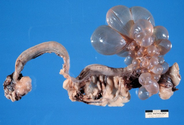
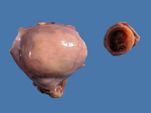
Dow (1957), when describing the complex called the cystic hyperplasia-pyometra complex found 23 of 100 dogs with CEH but no inflammation. Six of these had E. coli recovered from their uteri, indicating a subclinical infection. There were also 17 animals with CEH, but they had plasma cells in the stroma. Antigen recognition was occurring, but the source of the antigen was not known. Subclinical infection is possible. Dow 
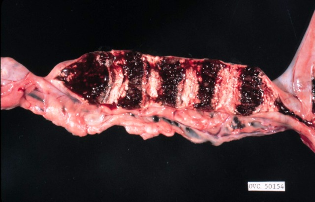
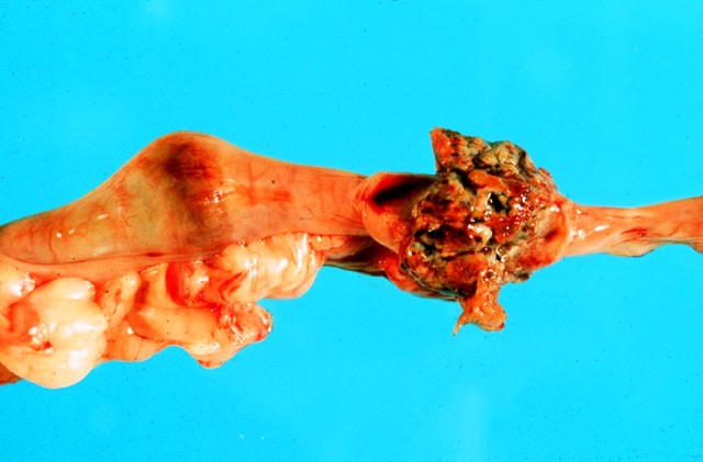
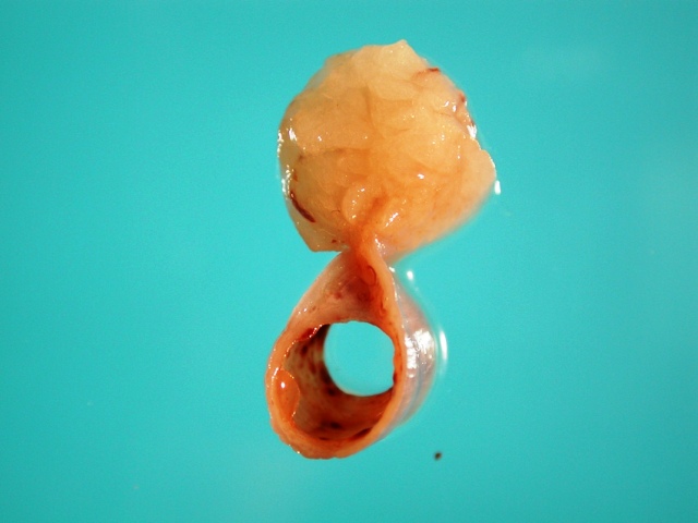
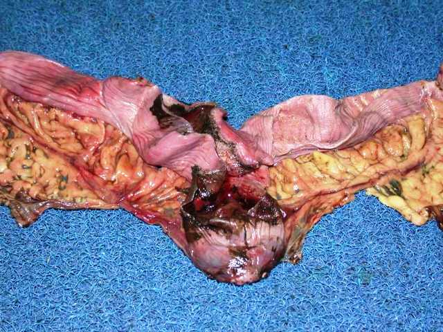
had 49 bitches with obvioius endometritis and pyometra. These had a variable amount of pus in the lumen, the amount varying with the degree of patency of the cervix. Three were unilateral. No bacteria were found in five cases, E coli was isolated in all but 4 of the remainder. Staphlococci and streptococci were isolated from a small number. Fourteen cases were called chronic endometritis. Those with 
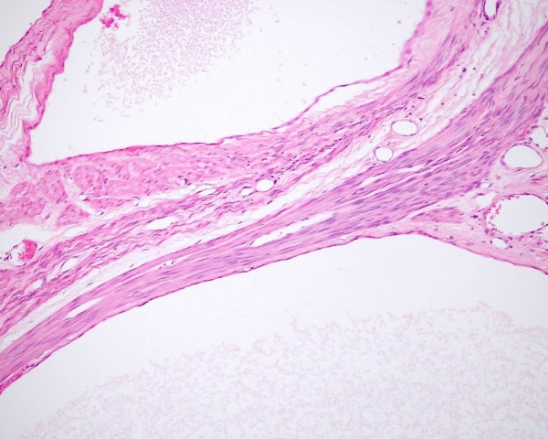
a closed cervix had massive dilation of the uterus, and a wall that was very atrophic and glands were often absent and the surface had squamous metaplasia. When the cervix was open, the endometrium was atrophic and glands were rare. There was fibrosis and numerous lymphocytes and plasma cell were abundant. In 1958, Dow reported additional cases 300, and they were categorised in a similar way. Results were similar. In 1959 Dow (1959) used the hormones estrogen and progesterone to induce CEH and endometritis in ovarectomised dogs. Dogs given cycles of estrogen followed by progesterone developed CEH and endometritis. The endometritis was dominated by neutrophils in the stroma and epithelium, and when progesterone was withdrawn, there was a change to plasma cell accummulation, and to resolution of inflammation. This occurred in cases where the uterus was isolated by a ligature around the distal part of a horn. The reaction was identical to that see when no ligature was present. Endometritis was clearly associated with the presence of progesterone - especially the neutrophilic response. Plasma cells dominated in uteri of dogs when progesterone therapy was stopped. In most cases, E coli was isolated from inflammed uteri.

Hadley and Osbourne (1974) reviewed the pathophysiology of pyometra. They recognised hormonal and bacterial factors. Pyometra occurs in dioestrus when the uterus is influenced by progesterone. Progesterone causes endometrial proliferation and secretion, and provides an environment for bacterial proliferation. They considered that bacteria were secondary invaders that were not necessary for the development of pyometra and therefore complicate the disease, rather than initiated it!

Hadley (1975) found that the serum oestrogen and progesterone concentration was within normal limits in single samples collected from dogs with false pregnancy and in dogs with pyometra.

Nomura (1983) induced endometritis experimentally by infusing the uterus with E coli from a case of pyometra in dogs at oestrus. Arora et al (2006) ovarectomised dogs and used one cycle of hormones and infused Escherichia coli to experimentally induce pyometra.

De Bosschere et al (2002) found that in endometritis-pyometra, estrogen receptors (ER) were lower in expression and progesterone receptors (PR) were higher in expression than in cystic endometrial hyperplasia, suggesting that ER and PR expression was different and that the pathogenesis was different. They suggested the presence of bacteria was the most important factor, rather than the widely held view that endocrine factors were the primary factors and bacterial infection was secondary (Kivisto et al 1977)

It is known that dogs with pyometra due to E coli develop anti E coli anitbody specific to the strain of E coli that infects the individual uterus (Kivisto et al 1977, Borresen and Naess 1977). What part these play in the pathogenesis is not known.

Schlafer and Gifford (2008) reviewed cystic endometrial hyperplasia and pyometra.

Maddens et al (2010) evaluated renal function in bitches with pyometra with E coli and identifed that the clinical signs of renal dysfunction were the result of a transient glomerular and proximal tubular dysfunction.

Bartoskova et al (2012) examined the number and percentages of B cells, T cells, T-helper cells, cytotoxic T cells and gamma delta T cellsin the uterine of normal dogs and those with pyometra. The percentages and ratios of B and T cells did not change but there were was a marked increase in the number of gamma delta T cells in pyometra. CD8- gamma delta T cells were particularly prominent. Neutrophils were the dominant cell population. There was a regulation a pro-inflammatory cytokines and down-regulation of inhibitory cytokines.

England et al (2012) studied normal dogs and those affected with endometrial hyperplasia examining mating induced endometrial inflammation (which is predominantly neutrophilic) and the attachment of spermatozoa to the epithelium. They found that mating induced endometritis occurs in bitches, and there is reduced adherence of spermatozoa to the endometrium of bitches with endometrial hyperplasia.

Yasunaga et al ( 2013) examined the vaginal and uterine mucosa for the presence of glucose and mannose using lectins and found that dogs with pyometra had altered expression during days 7-10 adn 30-40 of diestrus. They surmised this variance was linked to the susceptibility of some dogs to develop pyometra.

Holsta et al (2013) recorded alterations in the function of phagocytes in the later phases of dioestrus that may contribute to pyometra.


Macroscopic changes

Pyometra is usually a bilateral disease and affects the entire uterus including both uterine horns and uterine body. Unilateral pyometra is reported (Whitney 1969). Whitney (1969) reported 12 cases, only one of which was a true unilateral lesion and it was determined that the apparent unilateral nature was the result of interference with drainage from one side. There is often an irregular saccular distension of the uterus, but uniform distension is also seen.

The consistent finding in pyometra is mucopurulent material within the lumen of the uterus. The lumen is dilated to a variable extent and in extreme cases up to 6 cm diameter. Most are about 2 cm diameter.

Endometrial hyperplasia is usually, but not always, present.

Endometrial hyperplasia is invariably present in cases of endometritis and pyometra, but the severity of the hyperplasia varies (Misirlioglu et al 2006). The macroscopic changes of endometritis are similar to those of endometrial hyperplasia, with the exception that there may be an exudate reassembling pus. When pyometra is present, the lumen of the uterus is distended and filled with pus. This can be up to 1.5 litres!

Microscopic changes

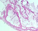
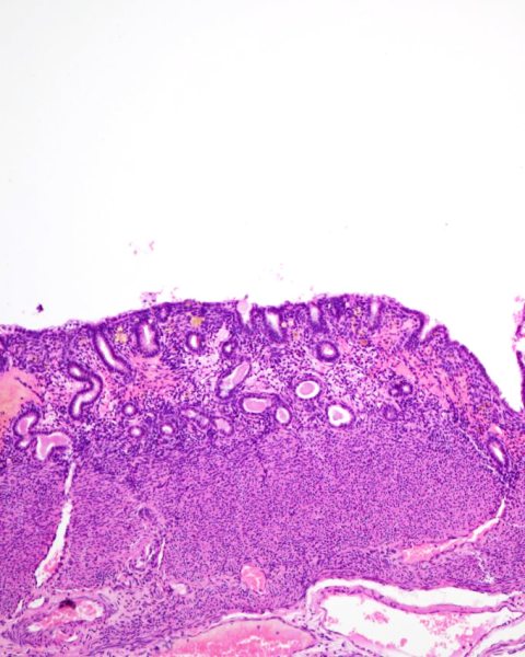
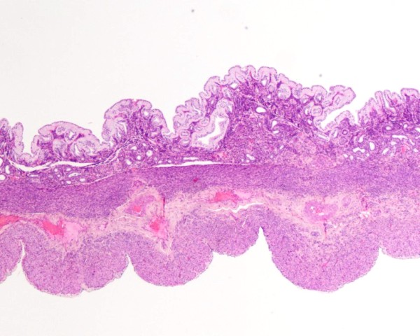
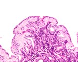
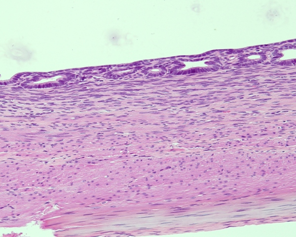
In most cases of endometritis, the histological lesions begin with a migration of neutrophilsand sometimes eosinophils into the glands and the lumen. Lymphocytes and plasma cells will form in the stroma of the endometrium. With a greater severity, particularly if the bacteria are toxigenic, there will be erosion and ulceration of the endometrium. Some cases just continue to accumulate neutrophils and the lumen will be tremendously dilated. The endometrium will not be so severely affected, and in some animals, squamous metaplasia will develop. The wall may become atrophic.

While neutrophils predominate, some cases have large numbers of granulated polymorphonuclear cells that resemble eosinophils.

In endometritis caused by Brucella canis the lesion is of a lymphohistiocytic type (Brennan et al 2008).

Mir et al (2013) reported on the findings of 21 bitches with either not confirmed pregnancy or unexpected pregnancy loss biopsied by laparotomy. Six of the 11 bitches with lesions and endometrial fibrosis, four had endometritis and to had cystic endometrial hyperplasia. Two had localised endometrial hyperplasia (PEH). A mixture of lesions were seen in 4 of the 11 with histological lesions.No bitches had bacteria recovered from the uterus.

Gifford at al (2014) reported on 399 cases where some fertile bitches had full thickness endometrial biopsies. They reported on the full range of lesions present in the uterus. 170 (43%) of cases had endometritis. Of these one half (52%) had a predominantly lymphocytic or lymphocytic and plasmacytic cell population predominating, 30% had a combination of neutrophils, lymphocytes and plasma cells, and the remaining 18% had predominantly neutrophils, eosinophils (7%) or both. Of the 399 cases, 101 or 25% had fibrosis. Cystic endometrial hyperplasia was identified in 133 of the 399 (33%). As yet the likely breeding outcome of dogs with various types of endometritis and fibrosis has not been determined.


Arora N, Sandford J, Browning GF, Sandy JR, Wright PJ.(2006) A model for cystic endometrial hyperplasia/pyometra complex in the bitch.Theriogenology. 66(6-7): 1530-1536.

Baba E, Hata H, Fukata T, Arakawa A (1983). Vaginal and uterine microflora of adult dogs. Amer J Vet Res 44: 606-609.

Bartoskova A, Turanek-Knotigova P, Matiasovic J, Oreskovic Z, Vicenova M, Stepanova H, Ondrackova P, Vitasek R, Leva L, Moore PF, Faldyna M. (2012) Gamma delta T lymphocytes are recruited into the inflamed uterus of bitches suffering from pyometra. The Vet J 2012 194: 303-309

Borresen B, Naess B (1977) Microbial, immunological and toxicological aspects of canine pyometra.Acta Vet Scand 18: 569-571

Brennan SJ, Ngeleka M, Philibert HM, Forbes LB, and Allen AL (2008) Canine brucellosis in a Saskatchewan kennel. Can Vet J. 49(7): 703-708.

De Bosschere H, Ducatelle R, Vermeirsch H, Simoens P, Coryn M (2002) Estrogen and progesterone receptor expression in cystic endometrial hyperplasia and pyometra in the bitch. Animal Reprod Sc 70: 251-259

Dow C (1957). The cystic hyperplasia-pyometra complex in the bitch. Vet Rec 69: 1409-1415

Dow C (1958) The cystic hyperplasia-pyometra complex in the bitch. Vet Rec 70: 1102-1109

Enginler SO, Ate A, Diren Sıgırcı, B, Sontas BH, Sonmez K, Karacam E, Ekici H, Evkuran Dal G, Gurel A. (2014) Measurement of C-reactive protein and Prostaglandin F2a Metabolite Concentrations in Differentiation of Canine Pyometra and Cystic Endometrial hyperplasia/ Mucometra. Reprod Dom Anim 2014 49: 641-647Dow C (1959) Experimental reproduction of the cystic hyperplasia-pyometra complex in the bitch. J Pathol Bacteriol 78: 267-278.

England GCW, Burgess CM, Freeman SL. (2012) Perturbed sperm–epithelial interaction in bitches with mating-induced endometritis. The Vet J 2012, 194: 314-318.

England GCW, Russo M, Freeman S (2013) The bitch uterine response to semen deposition and its modification by male accessory gland secretions. The Vet J 2003, 195: 179-184

Fransson B, Lagerstedt AS, Hellmen E, Jonsson P. (1997) Bacteriological findings, blood chemistry profile and plasma endotoxin levels in bitches with pyometra or other uterine diseases. Zentralbl Veterinarmed A. 44(7): 417-426

Gibson A, Dean R, Yates D, Stavisky J (2013) A retrospective study of pyometra at five RSPCA hospitals in the UK: 1728 cases from 2006 to 2011 Vet Rec 2013; 173: 396

Gifford AT, Scarlett JM, Schlafer DH. Histopathologic findings in uterine biopsy samples from subfertile bitches: 399 cases (1990-2005). J Am Vet Med Assoc 2014; 244: 180-186

Gogny A, Bruyas JF, Fiéni F. Pyometra in an inguinal hernia in a bitch. Reprod Domest Anim. 2010; 45: e461-464.

Hadley RM, Osbourne CA (1974) Canine pyometra: pathophysiology, diagnosis and treatment of uterine and extrauterine lesions. J Amer Anim Hosp Assoc 10: 245-268

Hadley JC (1975) Unconjugated oestrogen and progesterone concentrations in the blood of bitches with false pregnancy and pyometra. Vet Rec 96: 545-547

Hadley JC. (1975) The development of cystic endometrial hyperplasia in the bitch following serial uterine biopsies. J Small Anim Pract 16: 249-257.

Hardy RM, Osbourne CA (1974). Canine pyometra: pathophysiology, diagnosis and treatment of uterine and extra-uterine lesions. J Amer Anim Hosp Assoc 10: 245-268.

Veterinary Immunology and Immunopathology

Holst BS, Gustavsson MH, Lilliehöök I, Morrison D, Johannisson A (2013) Leucocyte phagocytosis during the luteal phase in bitches. Vet Immunol Immunopathol 2013, 153: 77-82

Ishiguro K, Baba E, Torii R, Tamada H, Kawate N, Hatoya S, Wijewardana V, Kumagai D, Sugiura K, Sawada T, Inaba T. (2006) Reduction of mucin-1 gene expression associated with increased Escherichia coli adherence in the canine uterus in the early stage of dioestrus. Vet J. in press

Kivisto A-K, Vasenius H, Sandholm M.(1977) Laboratory diagnosis of canine pyometra. Acta Vet Scand 18: 308-315.

Noakes DE, Dhaliwal GK, England GCW (2001) Cystic endometrial hyperplasia/pyometra in dogs: a review of the causes and pathogenesis. J Reprod Fert Suppl 57: 395-406

Maddens B, Daminet S, Smets P, Meyer E (2010) Escherichia coli Pyometra Induces Transient Glomerular and Tubular Dysfunction in Dogs. J Vet Intern Med 2010 24:1263-1270

Maciel GS, Uscategui RR, de Almeida VT, Oliveira MEF, Feliciano MAR, Vicente WRR. (2014) Quantity of IL-2, IL-4, IL-10, INF-γ, TNF-α and KC-Like Cytokines in Serum of Bitches With Pyometra in Different Stages of Oestrous Cycle and Pregnancy.Reprod Dom Anim 2014; 49: 701-704

Mir F, Fontaine E, Albaric O, Greer M, Vannier F, Schlafer DH, Fontbonne A. Findings in uterine biopsies obtained by laparotomy from bitches with unexplained infertility or pregnancy loss: an observational study. Theriogenology. 2013; 79: 312-322.

Nomura K. (1983) Canine pyometra with cystic endometrial hyperplasia experimentally induced by E coli inoculation. Jpn J Vet Sci 45: 237-240.

Nomura K, Kawasoe K, Shimada Y. (1990) Histological observations of canine cystic endometrial hyperplasia induced by intrauterine scratching. Jpn J Vet Sci 52: 979-983

Nomura K. (1994) Iinduction of a deciduoma in the dog. J Vet Med Sci 56: 365-369.

Nomura K. (1995) Histological evaluation of canine deciduoma induced by silk suture. J Vet Med Sci 57: 9-16.

Pretzer SD (2008) Clinical presentation of canine pyometra and mucometra; a review. Theriogenology 70: 359-363.

Rubio A, Boyen F, Tas O, Kitshoff A, Polis I, Van Goethem B, de Rooster H. Bacterial colonization of the ovarian bursa in dogs with clinically suspected pyometra and in controls. Theriogenology 2014; 82: 966-971.

Sandholm M, Vasenius H, Kivisto A-K. (1975) Pathogenesis of canine pyometra. J Amer Vet Med Assoc 167: 1006-1010.

Schultheiss PC, Jones RL, Kesel ML, Olson PN. (1999) Normal bacterial flora in canine and feline uteri. J Vet Diagn Invest 11: 560-562

Sevelius E, Tidholm A, Thoren-Tolling K. (1990) Pyometra in the dog. J Amer Anim Hosp Assoc 26: 33-38.

Schlafer DH, Gifford AT. (2008) Cystic endometrial hyperplasia, pseudo-placentational endometrial hyperplasia, and other cystic conditions of the canine and feline uterus. Theriogenology 70: 349-358

Smith FO. (2006) Canine pyometra. Theriogenology. 66(3): 610-612.

Stone EA, Littman MP, Robertson JL, Bovee KC. (1988) Renal dysfunction in dogs with pyometra. J Amer Vet Med Assoc 193: 457-464.

Verstegen J, Dhaliwal G,Verstegen-Onclin K. (2008) Mucometra, cystic endometrial hyperplasia, and pyometra in the bitch: Advances in treatment and assessment of future reproductive success. Theriogenology 70 (3 ): 364 - 374

Whitney JC. (1969) The pathology of unilateral pyometra in the bitch. J Small Anim Pract 10: 223-230.

Subinvolution of placental sites

Subinvolution of placental sites is the failure of one or more placental sites to involute or disappear at the appropriate time after parturition. Sloughing of the placental site is complete by 9 weeks and complete normal involution takes up to 12 weeks (Al Bassam et al 1981). The term 'subinvolution of placental sites' was first used by Beck and McEntee (1965) in a single case report. Riser (1940) reports a single case that resembled subinvolution, but were called different things ('chorioepithelioma' ). Glenn (1968) detailed 32 cases of subinvolution, providing clinical, macroscopic and microscopic findings. Al Bassam et al (1981) reported on involution abnormalities in the post partum uterus of dogs and detailed the changes seen in subinvolution (20 cases). They also reported on invasion of the myometrium with trophoblasts.

Clinical findings

The clinical signs of subinvolution are remarkable similar, being persistent postparturient vaginal bleeding (Glenn 1968). Jourdan and Hill (1951) reported on a bitch that had severe anaemia and emaciation 2 months after welping. She had a sanguinous vaginal discharge (which they called metrorrhagia). Treatment with progesterone and supportative therapy for anaemia resulted in resolution. The genital tract was normal when she was spayed 3 months latter. Is is likely this was a case of subinvolution of placental sites, a common postpartum cause of bloody vaginal discharge. While they called it metrorrhagia, we prefer to exclude oestral and postpartum discharges in the definition of metrorrhagia.

Pathogenesis

The factors responsible for subinvolution are not known. A consistent feature is retention and invasion of trophoblast like cells into the underlying stroma and even into the myometrium. Bacterial infection and hormonal abnormalities were not thought to be involved (Al Bassam et al 1981).

Macroscopic findings

Sites of subinvolution are larger than normal sites at the same stage of gestation. Placental sites are recognised as regular descrete oval enlargements that are evenly spaced along the horns and often the body of the uterus. Sometimes there will be a nodule protruding from the serosal surface of the placental site, representing bulging of the endometrium through the myometrium. Such as site can rupture and cause abdominal bleeding. The sites in cross section have marked thickening of the endometrium with red brown exudate and tissue, and multiple gray areas dispersed throughout. (Beck and McEntee 1965, Glenn 1968, Al Bassam et al 1981). Subinvolution can affect one or more of the placental sites, and seldom all of them.


Normal placenta sites in a postpartum bitch

Subinvolution of a placental site.

Microscopic findings

Histological findings reflect the macroscopic changes with there being expansion of the endometrium. There is considerable variation within one placental site and between placental sites. Abundant eosinophilic material is attached to and extends from the endometrium into the lumen. This is mostly fibrin and haemorrhage. Beneath this is stromal tissue composed of fibrous tissue containing large vascular spaces. Mineralised foci are scattered throughout. Epithelium is present in areas and individual cells are swollen and markedly vacuolated. Macrophages and lymphocytes and plasma cells are present in variable numbers. At the base of the lesion are individual multinucleated or clusters of syncitial cells, called trophoblasts. They are usually adjacent to the vascular spaces. In many cases, these invaded the myometrium and extend to the serosa. Endometrial glands are distended, and often much more than normal.

The features that histologically distinguish subinvolution from normal placental sites are large amounts of collagen in the placental site after 12 weeks post partum, collagen down to or extending into the myometrium, marked dilation of endometrial glands in the placental site, and trophoblast like cells in the collagen masses in the areas of invasion (Al Bassam et al 1981).


Al Bassam MA, Thomson RG, O'Donnell L. (1981) Normal postpartum involution in the uterus in the dog. Can J Comp Med 1981; 45: 217-232.

Al Bassam MA, Thomson RG, O'Donnell L. Involution abnormalities in the postpartum uterus in the bitch. Vet Pathol 1981; 18: 208-218.

Beck AM, McEntee K. (1966) Subinvolution of placental sites in a postpartum bitch. A case report. Cornell Vet. 1966; 26: 269-277.

Fernández PE, et al. Characterisation of cytotrophoblastic-like cells present in subinvolutioned placental sites of the bitch. Histol Histopathol 1998;13:995-1000.

Glenn BL (1968) Subinvolution of placental sites in the bitch. 18th Gaines Veterinary Symposium , Gaines Dog Research Centre, White Plains, NY p 7-10

Jourdan RH, Hill HJ (1951) A case of canine metrorrhagia. J Amer Vet Med Assoc 118-119: 377

Riser WH (1940). Chorioepithelioma of the uterus of a dog. J Amer Vet Med Assoc 96: 271.


Adenomyosis

Adenomyosis of the uterus is the presence of endometrial tissue within the myometrium. In animals apart from primates, the term endometriosis is not used. Adenomyosis of the uterus is often an incidental finding, but it can become a clinical condition if it is estensive or if the endometrial tissue, usually glands, becomes filled with secretion or exudate, bulges onto the serosa and ruptures. Peritonitis will ensue.

The pathogenesis is unknown, and there is speculation as to how the endometrium comes to be within the myometrium. According to Bloom (1954), serial section of affected regions of the uterus show a connection to the endometrium. Endometrial hyperplasia is usually present. Some assume the endometrium 'grows' into the myometrium, but the stimulus or cause for such an invasion is not clear. Likewise, being forced into the myometrium under pressure is equally difficult to imagine. Having said this, McEntee (1990) reports seeing an artefact of fixation where endometrium appears in the myometrium in uteri fixed without being opened, presumably due to contraction of the myometrium. He considered it essential to open the uterus prior to fixation to prevent endometrium being forced into the myometrium (McEntee 1990).

Adenomyosis is well recognised in human females where there is an association with increasing parity, early menarche (<10 years of age), short menstrual periods (<24 days), obesity and use of estrogenic hormones (Templeton et al 2007). One theory of the formation of adenomyosis (also called uterine endometriosis) is that adenomyosis results from hyperperistalsis or dysperistalsis of the uterus leading to increased intrauterine pressure and myometrial dehiscence and subsequent invasion of the endometrium into the myometrium. Thus there is mechanical trauma involved in its pathogenesis. (Leyendecker et al 2004).

Stocklin-Gautschi et al (2001) report on 2 cases of focal uterine adenomyosis and is one of the only reports of this disease. This may be because it is so common and of little clinical significance in itself to have warranted attention. In the 2 cases they report, the clinical signs were referable to the lesions of adenomyosis. The first case had a lesion in the uterine body at the cervix. It was 4-5 cm in diameter. The second case had the lesion in a uterine horn and there was torsion of the horn. The lesion was 8 cm diameter. Their first case was identical to that of Tamada et al (2005).

I suspect Tamada et al (2005) in their report of adenomyosis of the cervix are probably looking at adenomyosis of the uterine body as the lesion they report was 2-3 cm with cysts up to 6 mm in diameter. The canine cervix is about 1 cm long and is composed of the genital muscular body and a central canal that is oriented dorsoventrally (Roszel 1992). Adenomyosis of this structure would produce an intraluminal structure and not the specimen they illustrate in their manuscript.

Schlafer and Gifford (2008) in their review describe how cases of adenomyosis can progress to the stage where the epithelium of the glands disappears and leaves cavities of fluid within the wall of the uterus.

Adenomyosis is reported in studies on uterine biopsy in bitches including those of Mir et al (2013), Gifford et al (2014).


Macroscopic findings

In most cases, the gross lesions of adenomyosis are difficult to recognise grossly. It appears as though adenomyosis often occurs in the uterine body, and if severe can cause a thickening of that region. The thickness of the uterus can approach 8 cm diameter. When the lesion is this dramatic, the uterine wall is white and tough indicating fibrous tissue, and there will be small cystic structures visible within. These will contain exudate if there is concommitent pyometra.

Microscopic findings

Some cases of adenomyosis are identified only by the very observant, and even then, must be differentiated from sectioning artefact. Where the amount of endometrial tissue in the myometrium is extensive, the diagnosis is made easier. The endometrial tissue within the myometrium includes epithelium and stroma, and there is often hyperplasia of the elements, just like the adjacent endometrium. Cystic dilation of the epithelial components is common.

Roszel JF (1992) Anatomy of the canine uterine cervix. Compend Contin Edu 14: 751-760

Leyendecker G, Kunz G, Herbertz M, Beil D, Huppert P, Mall G, Kissler S, Noe M, Wildt L (2004) Uterine peristaltic activity and the development of endometriosis. Ann N Y Acad Sci. 1034: 338-355.

Schlafer DH, Gifford AT. (2008) Cystic endometrial hyperplasia, pseudo-placentational endometrial hyperplasia, and other cystic conditions of the canine and feline uterus. Theriogenology 70: 349-358

Stocklin-Gautschi NM, Guscetti F, Reichler IM, Geissbuhler U, Braun SA, Arnold S. (2001) Identification of focal adenomyosis as a uterine lesion in two dogs. J Small Anim Pract 42(8): 413-416.

Tamada H, Kawate N, Inaba T, Kuwamura M, Maeda M, Kajikawa T, Sawada T. (2005) Adenomyosis with severe inflammation in the uterine cervix in a dog. Can Vet J. 46(4): 333-334.

Templeman C, Marshall SF, Ursin G, Horn-Ross PL, Clarke CA, Allen M, Deapen D, Ziogas A, Reynolds P, Cress R, Hoda Anton-Culver H, West D, Ross RK, Bernstein L(2007) Adenomyosis and endometriosis in the California Teachers Study. Fertility and Sterility 90: 415-424

Endometriosis and endometrioma

Domestic mammals dont get endometriosis; only primates do. This is dogma. Yet people try to diagnose endometriosis. The intrauterine form in humans has a corelate in animals - we call it adenomyosis (above).

So how about the report of endometrioma by Paiva et al (2015)? They report a case of a 20cm mass near the uterus that was composed of tissue that they believe to be endometrial tissue. The gross appearance is reminiscent of a uterine polyp. Could this be a ruptured uterus with external polyp?

Paiva BH, Silva JF, Ocarino NM, Oliveira CA, Assis WA, Serakides R. A rare case of endometrioma in a bitch. Acta Vet Scand. 2015 Jun 18;57:31


Mucometra/hydrometra

The accumulation of serous fluid within the uterus is hydrometra, and if the fluid is mucinous, it becomes mucometra. The contents in these conditions are acellular or poorly cellular. It is normal for there to be increased fluid in the uterus during oestrus, and in cystic hyperplasia. Hydrometra or mucometra is a common part of pseudopregnancy (or localised endometrial hyperplasia).

In most conditions, the accumulation of fluid is the result of an obstruction to outflow. Cystic endometrial hyperplasia can contribute to this, and most cases of CEH have some degree of mucometra. Obstruction to outflow is seen in improper attempts to remove the uterus during ovariohysterectomy and leaving a portion of uterus behind (see ovarian remnant syndrome). The uterine remnant becomes cystically distended with clear fluid.

Obstruction at the cervix (physiological closure or lack of opening/estrus) will cause the uterine body and both horns to distend. Endometrial polyps can cause a local obstruction and the uterus will distend proximal to the site. Segmental aplasia and uterine neoplasia are other potential causes. Those cases where obstruction is the cause would be expected to have atrophy of the endometrium due to pressure.

Hydrometra and mucometra to a mild degree are also seen in cystic endometrial hyperplasia of its different forms, and varying degrees of dilation may be seen. It is also a part of pseudopregnancy. In mild cases, the dilation is best recognised after transverse sectioning of the uterus (see cystic endometrial hyperplasia above). In cystic endometrial hyperplasia due to progesterone stimulation, there is increases secretion by the endometrial glands, decreases myometrial contractility and causes closure of the cervix.

Marked dilation with hydrometra will normally require some obstructive situation. Oh et al (2005) for example found hydrometra in a dog with segmental aplasia of the uterine body. Pena et al (2006) reports mucometra in a dog with a large uterine carcinoma.

Cases of mucometra or hydrometra should be sterile (Fransson et al 1997).

One of the early reports of hydrometra was by McAfee and McAfee (1976) wherein they describe a young dog with 240 ml of serosanguinous fluid in the left uterine horn. The right horn and body were normal. There were 'localized areas of endometrial hypoplasia'.

Payan-Carreira et al (2006) reports on a case of hydrometra in a 10 yr old Yorkshire terrier where there was no endometrial hyperplasia or obstruction, yet the uterus was 2 cm diameter. There was a microscopic granulosa cell tumour, but no corpora lutea or follicles. There was no endometrial hyperplasia but there was widespread estrogen receptor reactivity on immunohistochemistry. They surmised the granulosa cell tumour was responsible for stimulating this lesion - oestrogen production unregulating oestrogen receptor activity in the uterus.

Sontas et al (2013) reported on a dog with inguinal hernia that contained the uterus - which was reported to have hydrometra/mucometra. The mucometra/hydrometra appeared to be very mild. The histological finding of endometrial fibrosis could be from entrapment. Hemorrhage and edema of the endometrium was reported - how this differs from an identical finding from surgical manipulation was not discussed.

Fransson B, Lagerstedt AS, Hellmen E, Jonsson P. (1997) Bacteriological findings, blood chemistry profile and plasma endotoxin levels in bitches with pyometra or other uterine diseases. Zentralbl Veterinarmed A. 44(7): 417-426

Mucometra is reported in studies on uterine biopsy is in bitches including those of Mir et al (2013) and Gifford et al (2014)


.McAfee LT, McAfee JT (1976) Hydrometra in a bitch. Mod Vet Pract 57: 829.

McEntee K (1990) Reproductive Pathology of Domestic Mammals. Academic Press p 170.

Oh KS, Son CH, Kim BS, Hwang SS, Kim YJ, Park SJ, Jeong JH, Jeong C, Park SH, Cho KO. (2005) Segmental aplasia of uterine body in an adult mixed breed dog. J Vet Diagn Invest. 17(5): 490-492.

Payan-Carreira R, Pina J, Costa M, Seixas F, Pires MA. (2006) Oestrogen receptors in a case of hydrometra in a bitch. Vet Rec. 158(14) :487-489

Pena FJ, Gines JA, Duque J, Vieitez V, Matrinez-Perez R, Madejon L, Nunez Martinez I, Moran JM, Fernandez-Garcia S (2006). Endometrial Adenocarcinoma and Mucometra in a 6-year-old Alaska Malamute. Reprod Dom Anim. 41: 189-190.

Pretzer SD (2008) Clinical presentation of canine pyometra and mucometra; a review. Theriogenology 70: 359-363.

Sontas HB, Toydemir S, Erdogan O, Sennazli G, Ekici H (2013) Inguinal herniation with hydrometra/mucometra in a poodle bitch. Canadian vet J 2013; 54: 840-844.


Macroscopic findings.

The affected uterine horn (s) and or body are of a larger diameter than normal and their contents are clear fluid (hydrometra) or mucus (mucometra). The lumen is dilated and the contents will empty. There should be some indication of an obstruction, especially if the dilation is obvioius. In severe cases, the wall will be thin and stretched.

Microscopic findings.

The histological changes reflect the macroscopic changes. While many people concentrate on the wall of the uterus, the presence of dilation of the viscus is often overlooked. In severe cases, the wall will be thin. The endometrium may be atrophic and the glands reduced to tiny depressions or outpouching of the luminal epithelium. In less severely affected examples, there may be some endometrial hyperplasia, and especially in mucometra, the epithelium may have a decidual reaction - abitch in diestrus will have hypertrophy of the luminal epithelial cells such that they are tall columnar and have a highly vacuolated cytoplasm.

Hydrometra in a bitch. The uterine wall is thin and the lumen is dilated (YB196569)

Mucometra in a bitch. The wall is thin, and in this case their is a decidual reation of the luminal epithelium (YB106871)


Cysts and embryonic remnants

The section on remnants of the mesonephric and paramesonephric systems for the ovary and uterine tube apply here as well. In the area of the uterus, there are several choices for cysts. They are:-

  • cystic remnant of mesonephric duct
  • cystic remnant of the paramesonephric duct or uterine duplication
  • cystic adenomyosis (see adenomyosis)
  • serosal inclusion cysts
  • endometrial cysts in cystic endometrial hyperplasia (see cystic endometrial hyperplasia).

Embryonic remnants and duplications (mesonephric and paramesonephric ducts)

The most common location for these are in the mesometrium dorsal to the uterine horns and bilaterally on each side of the uterine body. They continue on at the lateral sides of the vagina to become Gartners ducts (see Vagina and Vulva).

Animals with disorders of sexual development such as those with 'male pseudohermaproditism or feminisation' have remnants of the mesonephric ducts that are histologically identical to the deferent ducts (which are derived from the mesonephric ducts) that run dorsal to the uterus in the mesometrium and then lateral to the uterine body. It is not uncommon for these to be present in otherwise normal females, but typically there are only segments of the ducts. In some cases these are incidental structures seen histologically, but in some they form a cyst or a mass that prompts the practitioner/surgeon to submit them for histological evaluation. When the structures are similar to a cystic deferent duct with a simple columnar epithelial lining and a complete wall of smooth muscle, there is little challenge and the diagnosis of remnant of the mesonephric duct can be made.

Occasionally I see cystic structures in the location of the remnants of the mesonephric ducts that have an inner lining of endometrium. I have even seen epithelial hypertrophy and vacuolation that is typical of dioestral endometrium, and some with endometrial glands! In these cases I have little choice but to call them cystic remnants of paramesonephric ducts or uterine duplication. I suspect these are pluropotential structures and could be either mesonephric or paranephric in appearance. After all, both structures are formed by invagination of coelomic tissue (or tissues destined to be mesothelium).

Recently, Bartel et al (2011) published 3 cases that they called ectopic endometrium in mesonephric remnants of the uterus. They used immunohistochemistry for hormone receptors, cytokeratin and smooth muscle actin to show the lining of the tubules was endometrium, and that the remnants were in the typical location of the mesenephic tubules. Considering the embryology of the ducts and the appearance, I would not call this ectopia but differentiation, and because differentiation and phenotype determine the names we give, it seems unduly complicated to call this 'Ectopic Endometrial Tissue in Mesonephric Remnants' especially when they did not prove the ducts were truely mesonephric!

I have seen many of these and at least 10 are recorded up to 2006.

Cystic remnant of mesonephric duct dorsal to the uterus of a dog.

Histology of the wall of a cystic remnant of mesonephric duct in a dog (YB165919)

Bartel C, Berghold P, Walter I. (2011) Ectopic Endometrial Tissue in Mesonephric Duct Remnants in Bitches. Reprod Domestic Animal 2011

McEntee (1990) reports seeing duplication of the uterine horn in a cat and a dog, and published a photo of a canine example.

Schlafer and Gifford (2008) speak about this and provide a photograph too.


McEntee K (1990) Reproductive Pathology of Domestic Mammals. Academic Press p 121-122.

Schlafer DH, Gifford AT. (2008) Cystic endometrial hyperplasia, pseudo-placentational endometrial hyperplasia, and other cystic conditions of the canine and feline uterus. Theriogenology 70: 349-358

Serosal inclusion cysts

Serosal inclusion cysts are common in dogs, but they are very rare in cats. They are thin walled fluid filled cystic structures on the serosal surface of the uterus. They are often multilocular and are attached by a thin stalk. They have a lining of thin squamous (flattened) cells that in some parts of the cyst may become cuboidal. The wall is composed of compressed collagen. They are believed to arise when mesothelial cells become trapped in the serosa and continue to secrete, become hyperplastic and form the characteristic structures.

McEntee (1990) believed they develop during post partum uterine involution.

Arnold et al (1996) wrote a case report of one case and detailed the ultrasonographic, macroscopic and histological appearance.

Ortega-Pacheco et al (2006) examined 300 stray bitches and found that 5% had serosal inclusion cysts.

In their review, Schlafer and Gifford 92008) reiterate the suggested pathogenesis of serosal inclusion cysts.

There are 17 cases in the YagerBest Histovet database.

.....

Serosal inclusion cysts that project as grape like clusters from the serosa of the uterus.

Histology of multiple serosal inclusion cysts. Each cyst is lined by a simple low cuboidal layer of mesothelium (YB159544).


Arnold S, Hubler M, Hauser B, Kaser-Hotz B, Rusch P. (1996) Uterine serosal inclusion cysts in a bitch. J Small Anim Pract 37(5): 235-237.

McEntee K (1990) Reproductive Pathology of Domestic Mammals. Academic Press p 158-159

Ortega-Pacheco A, Segura-Correa JC, Jimenez-Coello M, Linde Forsberg C. (2006) Reproductive patterns and reproductive pathologies of stray bitches in the tropics. Theriogenology.

Schlafer DH, Gifford AT. (2008) Cystic endometrial hyperplasia, pseudo-placentational endometrial hyperplasia, and other cystic conditions of the canine and feline uterus. Theriogenology 70: 349-358


Uterine Neoplasia

Traditional classification schemes of neoplastic disease often focus on whether the pattern of differentiation is epithelial, mesenchymal, round cell or other. This type of classification works well in the uterus, however, distribution of the lesions is an all important factor, so this is the approach used here.

Myometrial neoplasia

Smooth muscle tumors (leiomyoma, fibroleiomyoma, fibromyxoleiomyoma, adipoleiomyoma, leiomyosarcoma)

The most common neoplasm of the wall of the uterus in the dog is the smooth muscle tumor (leiomyoma or leiomyosarcoma). These diagnoses are not based on clinical outcome. This is well known and I personally have seen hundreds of these with very few identified as metastatic. In fact there was one metastatic case I saw and it was classified histologically as a leiomyoma. Avallone et al (2022) stated "There was no statically significant correlation between diagnosis
and relapse or death from tumor-related causes."

I no longer classify smooth muscle tumors of the uterus as leiomyoma or leiomyosarcoma or smooth muscle tumor of unknown metastatic potential. I just call them smooth muscle tumor.

The most common smooth muscle tumor of the uterus has traditionally been classified as leiomyoma. When the neoplasm has a abundant fibrous tissue with the smooth muscle, they are sometimes called fibroleiomyoma. Other types of mesenchymal tissue may be present and the name reflects the amount of this other tissue.

Immunistochemistry should show these to be smooth muscle actin positive, calponin positive, as well as vimentin positive, although in some smooth muscle tumours, vimentin can be negative. It is worthwhile being on the lookout for the CD117 (cKit) + stromal tumour called gastrointestinal stromal tumour, which is usually found in the intestine, but which in humans is occasionally found in the reproductive tract.

In dogs, the criteria to classify a smooth muscle tumour as malignant is not defined with any kind of precision. It is not based on any studies of clinical outcome.

In humans, only those with a high probability of metastasis are designated as leiomyosarcomas. In humans, leiomyosarcoma is diagnosed when there is

    • mitotic count > 15 p10HPF and marked nuclear atypia and hypercellularity,
    • and supported by invasion, extrauterine extension, size over 10 cm, and tumor necrosis.

Thus a diagnosis of leiomyosarcoma in humans carries a very poor prognosis.

Those tumors that do not fulfill these criteria but have atypical histologic features are called smooth muscle tumors of uncertain malignant potential (STUMP) or atypical leiomyomas. All the rest are called leiomyomas.

If we apply these criteria to smooth muscle tumors of the uterus in animals, there would be virtually no leiomyosarcomas. In addition, reports of metastatic smooth muscle tumors are exceptionally rare in animals.

Avallone G, Pellegrino V, Muscatello LV, Roccabianca P, Castellani G, Sala C, Tecilla M, Valenti P, Sarli G. Canine smooth muscle tumors: A clinicopathological study. Vet Pathol. 2022; 59: 244-255.


Leiomyoma (according to the literature)

Brody and Roszel (1967) report finding 10 leiomyomas of the uterus.

In their study of German Shepherd with nodular dermatofibrosis and multifocal renal cystadenocarcinomas, Lium and Moe (1985) reported multiple uterine leiomyomas in 10 of 11 bitches with nodular dermatofibrosis.

Andreasen and Mahaffey (1987) in their study of desmin expression of smooth muscle tumours examined 2 that were located in the uterus.

McEntee (1990) reports seeing leiomyomas in 27 dogs.

Suresh Kumar et al (1995) published a case report of a leiomyoma in the uterus of a bitch.

Millan et al (2007) reports finding 4 leiomyomas of the uterus. They had 28 cases of leiomyoma of the genital tract. Their publication examined steroid receptors in canine and human smooth muscle tumours, but the results of uterine tumours were not separated from the others..

Ozmen et al (2008) reported a single case of multiple leiomyomas in a bitch - they were in the uterus, cervix and vagina. Immunohistochemical staining was done - they are muscle specific actin positive, pancytokeratin and S100 negative, desmin was slightly positive.


Andreasen CB, Mahaffey EA. (1987) Immunohistochemical demonstration of desmin in canine smooth muscle tumors. Vet Pathol 24(3): 211-215.

Avallone G, Pellegrino V, Muscatello LV, Roccabianca P, Castellani G, Sala C, Tecilla M, Valenti P, Sarli G. Canine smooth muscle tumors: A clinicopathological study. Vet Pathol. 2022; 59: 244-255.

Brody RS, Roszel JF (1967) Neoplasms of the canine uterus, vagina and vulva: a clinicopathologic survey of 90 cases. J Amer Vet med Assoc 151: 1294-1307.

D'Angelo E, Prat J.  Uterine sarcomas: A review. Gynecol Oncol. 2010;116:131-139.

Lium B, Moe L.(1985) Hereditary multifocal renal cystadenocarcinomas and nodular dermatofibrosis in the German shepherd dog: macroscopic and histopathologic changes. Vet Pathol. 22(5): 447-455.

McEntee K (1990) Reproductive Pathology of Domestic Mammals. Academic Press p 143.

Millan Y, Gordon A, Espinosa de los Monteros A, Reymundo C, Martin de las Mulas J. (2007) Steroid receptors in canine and human female genital tract tumours with smooth muscle differentiation. J Comp Pathol 136: 197-201

Ozmen O, Haligur M, Kocamuftuoglu M. (2008) Clinocopathologic and Immunohistochemical Findings of Multiple Genital Leiomyomas and Mammary Adenocarcinomas in a Bitch. Reprod Dom Animal. 43 (3): 377–381

Suresh Kumar RV, Ramakrishna O, Sreeraman PK. (1995) Leiomyoma uteri in a bitch. Can Vet J 36: 185.


Leiomyosarcoma(according to the literature)

Brody and Roszel (1967) report one case.

Tsioli et al (2011) reported one case of uterine leiomyosarcoma. The dog died 24 months later of unknown causes.

Avallone G, Pellegrino V, Muscatello LV, Roccabianca P, Castellani G, Sala C, Tecilla M, Valenti P, Sarli G. Canine smooth muscle tumors: A clinicopathological study. Vet Pathol. 2022; 59: 244-255.

Brodey RS, Roszel JF (1967) Neoplasms of the canine uterus, vagina and vulva: a clinicopathologic survey of 90 cases. J Amer Vet med Assoc 151: 1294-1307.

Tsioli VG, Gouletsou PG, Loukopoulos P, Zavlaris M, Galatos AD (2011) Uterine leiomyosarcoma and pyometra in a dog (pages 121–124) J Small Anim Pract 2011 52: 121-124

Angiolipoleiomyoma (or benign mesenchymoma?)

Boisclair and Dore (2001) report on a dog with a 10cm diameter mass composed mostly of mature adipose tissue with mature smooth muscle interspersed throughout. There were islands of cartilage and bone, and large tortuous arteries. Nomenclature in these unusual tumors can be confusing and because there is a variety of tissue, any combination of angio-, lipo-, leiomyo- or others would seem appropriate. The description of this mass also fits with a benign mesenchymoma.


Boisclair J, Dore M. (2001) Uterine angiolipoleiomyoma in a dog. Vet Pathol: 38: 726-728

Lipoleiomyoma

Percival et al (2018) described a single case of a giant lipoleiomyoma in a dog. It was 27x17x15 cm in a miniature poodle. This dog also had a leiomyoma too.


Percival A, Singh A, Zur Linden RA, Watrous G, Patten S, Valverde A, Ratsep E. Massive uterine lipoleiomyoma and leiomyoma in a miniature poodle bitch. Can Vet J. 2018; 59: 845-850.

Lipoma

Bloom (1954) reports seeing a lipoma in the mesometrium of a dog.


Endometrial neoplasia

The normal endometrium has epithelial and stromal components, so neoplasia involving epithelial only, stromal only or combinations are seen. All are rare, but epithelial tumours appear to be the more common. The features of each are similar to those see in other species.

Endometrial carcinoma

There are ocasional often single case reports of endometrial carcinoma in the dog. These reports are more numerous than other neoplasms of the endometrium and makes carcinoma the most likely neoplasm to be encountered.

Schlotthauer (1939) reports finding a adenocarcinoma of the uterus. It was a focal polypoid and cystic mass in the left horn of the uterus.

Joshi et al (1967) reported finding a uterine adenocarcinoma in a dog. The lesion was unilateral, involving one horn. The horn was twisted. The lesion was focal and there was invasion of epithelial cells into the myometrium. No metastases were present in other organs.

Koch and Kaiser (1976) report finding a multifocal adenocarcinoma of the uterus in a dog. It had extensive micrometastases throughout the body.

Vos (1988) reported finding 5 uterine and cervical carcinomas. All involved the uterus - 3 were multifocal and 2 involved the uterine horn and cervix. The three dogs with multifocal disease had metastases.

Payne-Johnson et al (1986) reported on a polypoid 3x1.5x1 cm mass of the uterine body that was a carcinoma.

Baldwin et al (1992) reviewed the literature up to 1992 and included a single case of uterine adenocarcinoma involving the uterine body. The neoplasm was multifocal in the body.

Cave et al (2002) reports on a single case. The dog was 10 months old and had a single large left uterine mass with surface ulceration. No metastases were present 2 years after ovariohysterectomy.

Pena et al (2006) reports a single case. The lesion was necrotic and mineralized and 30 cm diameter. It developed metastases in the lungs 9 months later.

Špoljarić B, et al. (2019) reported on a single case that they called a mucinous adenocarcinoma.

Pires et al (2019) reports a single case of a papillary mass that was invasive of the myometrium. The cells were PanCK and CK7 positive.


Immunohistochemistry of uterine adenocarcinoma

There are several antibodies used in evalution of the uterus including various cytokeratins, vimentin, smooth muscle actin and CD10. Staining is variable and not exactly consistent. Endometrial Adenocarcinoma is cytokeratin positive, and generally vimentin negative, CD10 positive, and smooth muscle actin negative.


Baldwin CJ, Roszel JF, Clark TP (1992) Uterine adenocarcinoma in dogs. Compend Contin Edu 14: 731-737

Cave TA, Hine R, Howie F, Thompson H, Argyle DJ. (2002) Uterine carcinoma in a 10-month-old golden retriever. J Small Anim Pract 43(3): 133-135.

Joshi KV, Sardeshpande PD, Jalnapurkar BV, Ajinkya SM (1967) A case of uterine adenocarcinoma in dog. Indian Vet J 44: 114-116.

Koch F, Kaiser E, (1976) Uteruscarcinom bei einem hund. Berliner und Munchener tierarztliche Wochenschrift. 89: 373-377.

Payne-Johnson CE, Kelly DF, Davies PT (1986) Endometrial carcinoma in a young dog. J Comp Path 96: 463-467.

Pena FJ, Gines JA, Duque J, Vieitez V, Matrinez-Perez R, Madejon L, Nunez Martinez I, Moran JM, Fernandez-Garcia S (2006). Endometrial Adenocarcinoma and Mucometra in a 6-year-old Alaska Malamute. Reprod Dom Anim. 41: 189-190.

Pires MDA, Catarino JC, Vilhena H, Faim S, Neves T, Freire A, Seixas F, Orge L, Payan-Carreira R. Co-existing monophasic teratoma and uterine adenocarcinoma in a female dog. Reprod Domest Anim. 2019l; 54: 1044-1049.

Schlotthauer CF (1939). Primary neoplasms in the genito-urinary System of dogs; a report of ten cases. J Amer Vet Med Assoc 95: 181-186

Špoljarić B, Vince S, Folnožić I, et al. Endometrial Mucinous Adenocarcinoma in a Bitch With Pyometra-A Case Report. Top Companion Anim Med. 2019; 36: 16-21.

Vos JH (1988) Uterine and cervical carcinoma in five dogs. J Vet Med 35: 385-390.


Endometrial stromal sarcoma (tumors)

In humans, 3 main entities are recognised - Endometrial stromal nodule, endometrial stromal sarcoma - low grade, and endometrial saroma - high grade.

Endometrial stromal sarcomas are very rare in dogs. They form a nodule of regional thickening of the endometrium and push aside, or focally invade the myometrium. Endometrial glands may be trapped within the neoplasm. They are stromal in type with spindle shaped cells in a myxomatous matrix that are arranged in a haphazard arrangement. Endometrial stromal nodule are well circumscribed, but sarcomas are infiltrative. These are called endometrial stromal sarcomas - low grade, and endometrial sarcoma - high grade.

I have only seen one convincing endometrial stromal tumour in a bitch. This case was a single lesion with a monomorphic population of cells that formed an infiltrative mass in the endometrium. The cells resembled smooth muscle, but were infiltrative and was restricted to the endometrium. This would have been an endometrial stromal sarcoma - low grade.

I have chosen to place the condition of endometrial polypoid adenomyomatosis with the hyperplastic lesions of the uterus. In the original report of this condition, the lesion was muliple and similar to a polyp with smooth muscle.

Endometrial stromal tumour in a bitch (YB156095).

Mixed endometrial tumours

Neoplasms of the endometrium with both epithelial and stromal elements are very unusual and rare. I read with interest the publication on endometrial polypoid adenomyomatosis (see above) where there were features that could be interpreted as a mixed endometrial tumour, except their example has multiple polyps.

I am unaware of any publications of mixed tumours in dogs.

Benign mixed tumour/adenoleiomyoma/benign mixed Muellerian tumour

I have seen on single large mass in the uterus of a dog that histologically was compatable with endometrial polypoid adenomyomatosis, but it was a single mass and it was 5 cm diameter. It was polypoid with a broad base and had epithelial and stromal components. It appeared to be benign both in its growth habit and histological appearance. Adenoleiomyoma is now my preferred diagnosis.

Adenoleiomyoma of the uterus in a dog. This single large mass resembled endometrial polypoid adenomyomatosis. Notice the glandular structures in the cross section (YB221758).

Carcinosarcoma (Malignant mixed tumour, malignant mixed Muellerian tumour)

Malignant mixed neoplasms of the endometrium have both epithelial and stromal elements, and either can be histologically benign or malignant. They are named according to the elements present and both should be anaplastic.

I have not seen a case in dogs

Other uterine neoplasia

Haemangiosarcoma

Murakai et al (2001) published a report of a 15 years old dog that developed abdominal enlargement from peritoneal metastases of a haemangiosarcoma. There was bilateral diffuse involvement of the uterus, which was massively enlarged. They believed the neoplasm originated in the uterus.

Murakami Y, Uchida K, Yamaguchi R, Tateyama S. (2001) Diffuse bilateral hemangiosarcoma of the uterus in a dog. J Vet Med Sci 63: 191-193.

Rhabdomyosarcoma

Bae et al (2007) reports a single case of a 2 yr old dog with metastatic rhabdomyosarcoma involving the bladder, retroperitoneal region and uterine wall, and metastases to multiple organs. Considering the involvement of the bladder, this may have begun as a typical bladder rhabdomyosarcoma and infiltrated the adjacent tissues including the uterus.

Bae I-H, Kim Y, Pakhrin B, . You M-H, Hwang C-Y, Kim J.-H, Kim D-Y. (2007) Genitourinary rhabdomyosarcoma with systemic metastasis in a young dog. Vet Pathol 44: 518-520

Plasmacytoma

Choi et al (2004) reported on an older dog with a mass in the uterus - the cervix to be specific. There was no disease after 10 months.

Choi YK, Lee JY, Kim DY, Park JI, Jeong SW, Park HM, Park C. Uterine extramedullary plasmacytoma in a dog. Vet Rec. 2004; 154: 699-700.

Neoplasia metastatic to uterus

Bastan et al (2008) report metstasis of canine transmissible venereal tumour to the uterus and ovaries of a 7 year old dog . This boxer had a vaginal CTVT.

Bastan A, Baki Acar D, Cengiz M (2008) Uterine and ovarian metastasis of transmissible venereal tumor in a bitch. Turk J Vet Anim Sci 32: 65-66

Placental neoplasia - Gestational trophoblastic Tumors

The umbrella term for tumors with a phenotype of placental tissue, predominantly trophoblasts is gestational trophoblastic disease. This includes neoplastic and nonneoplastic tumors. The nonneoplastic diseases are the placental or hydadiform moles, and the neoplasms are the gestational trophoblastic tumors, choriocarcinoma, placental site trophoblastic tumor, and epithelioid trophoblastic tumor.

Aggressive invasive trophoblasts (the dog equavalent of human placenta accreta spectrum) occur periodically. There is a review of the the human disease in the references.

Hecht JL, Baergen R, Ernst LM, Katzman PJ, Jacques SM, Jauniaux E, Khong TY, Metlay LA, Poder L, Qureshi F, Rabban JT 3rd, Roberts DJ, Shainker S, Heller DS. Classification and reporting guidelines for the pathology diagnosis of placenta accreta spectrum (PAS) disorders: recommendations from an expert panel. Mod Pathol. 2020; 33: 2382-2396.

Choriocarcinoma

Riser (1940) reported on a dog with 3 red nodules in the uterus, one in each horn and one at the bifurcation of the uterus. His diagnosis was chorioepithelioma, but several authors suggest this was actually subinvolution of placental sites with transmural invasion in one.

Christopher (1972) reports finding a choriocarcinoma in a bitch. It was an invasive 2x3 cm lesion in one horn in a bitch that had welped previously.

Poutahidis et al (2008) Reported on a primary gastric choriocarcinoma.


Christopher J (1972) Chorio-carcinoma in a bitch; a note. Indian J Anim Sc 42: 731-733

Poutahidis T, Doulberis M, Karamanavi E, Angelopoulou K, Koutinas CK, Papazoglou LG. Primary gastric choriocarcinoma in a dog. J Comp Pathol. 2008; 139: 146-150.

Riser WH (1940). Chorioepithelioma of the uterus of a dog. J Amer Vet Med Assoc 96: 271


Miscellaneous (rare and unusual) diseases of the uterus

Uterine calculi

Iwasaki and Oliveira (1991) report finding 2-3 mm white calculi within the uterus of a 7 yr old bitch. There was cystic endometrial hyperplasia and mucous was present in the lumen.

Iwasaki M, Oliveira CA. (1991) Uterine lithiasis in a dog. Aust Vet J 68: 73-74

Uterine haemorrhage (haematometra, metrorrhagia)

It is normal for the bitch to develop a small degree of 'haemorrhage' from the endometrium during oestrus. In the normal bitch there is 'extravasation' of red cells into the superficial endometrial stroma (McEntee 1990). There is a bloody vaginal discharge after parturition (lochia) but this is physiological and not included here. Subinvolution of placental sites is when there is prolonged bloody vaginal discharge after birth, and is also excluded, as is open pyometra.

Abnormal bleeding, not associated with oestrus or postpartum is called metrorrhagia. Uterine haemorrhage of this type is very rare and there are only a few reports of this in dogs. Most are due to a coagulopathy, and rodenticide anticoagulants are high on the list of possiblilities. Most are seen in young dogs after their first oestrus.

Rendano et al (1974) reports on one dog that 5 weeks after her first oestrus developed a haemorrhagic vulval discharge and blood loss anaemia due to marked haematometra.The uterus had marked intraluminal haemorrhage and haemorrhage into the wall and on the serosa. There were multiple sacculations and localised endometrial hyperplasia/deciduoma formation consistent the pseudopregnancy (pseudocyesis). The cause of the haemorrhage was not determined and a coagulopathy was considered. One horn of the uterus was twisted (torsion).

Wheeler et al (1984) reports on a dog with uterine and vaginal haemorrhage that had a factor VII deficiency.

Padgett et al (1998) reports on one 17 month old dog who presented with vaginal bleeding and anaemia. The dog had a coagulopathy from brodifacoum intoxication (anticoagulant rodenticide). The dog had haematometra.

Misumi et al (2002) report on a dog that had haematometria, and concurrent torsion of the uterus.

Troxel et al (2002) reported on a dog that had severe haemorrhage and it was eminating from the uterus. The underlying lesion was pyometra and endometrial hyperplasia.

Gumber et al (2010) reported on a dog with marked uterine hemorrhage originating from an endometrial polyp in a dog with open pyometra.

I have seen 2 cases of haemorrhagic shock of young bitches at oestrus - one dog had vonWillebrands disease and the other had toxicosis with anticoagulant rodenticide. The uterus in both cases was filled with blood and the dogs had bled profusely from the vulva.

There were 5 cases of 'idiopathic' uterine haemorrhage in the YagerBest Histovet database. These presumably had a coagulopathy.

Gumber S, Springer N, Wakamatsu N. (2010) Uterine endometrial polyp with severe hemorrhage and cystic endometrial
hyperplasia–pyometra complex in a dog. J Vet Diagn Invest 2010 22:455–458

McEntee K (1990) Reproductive Pathology of Domestic Mammals. Academic Press p 143.

Misumi K, Fujiki M, Miura N, Sakamoto H (2002) Uterine horn torsion in two non-gravid bitches. J Small Anim Pract. 2000 Oct;41(10):468-71

Padgett SL, Stokes JE, Tucker RL, Wheaton LG. (1998) Hematometra secondary to anticoagulant rodenticide toxicity. J Am Anim Hosp Assoc 34(5): 437-439.

RendanoVT, Juck FA, Binnington AG (1994) Hematometra associated with pseudocyesis and uterine torsion in a dog. J Amer Anim Hosp Assoc 10: 557-580.

Troxel MT, Cornetta AM, Pastor KF, Hartzband LE, Besancon MF (2002) Severe hematometra in a dog with cystic endometrial hyperplasia/pyometra complex. J Am Anim Hosp Assoc. 38(1): 85-89.

Wheeler SL, Weingand KW, Thrall MA, Berg RJ, Schwarz PD, Olson PN. (1984) Persistent uterine and vaginal hemorrhage in a beagle with factor VII deficiency. J Amer Vet Med Assoc 185: 447-448.

Uterine hernia

The uterus, as with any abdominal viscera, can herneate through the diaphragm, abdominal wall or through any opening. The uterus is usually gravid for this to occur.

Snow (1956) reports on herneation of the uterus into the inguinal region through the inguinal ring.

Sullivan et al (1969) and Bellenger et al (1975) both report on herneation of the gravid uterus into the chest.

McNamara et al (1997) report on herneation of the non gravid uterus into a vaginal cervical prolapse.

Sontas et al (2013) reported on a bitch with inguinal hernia that contained the uterus. The uterus was not pregnant. It had mild mucometra/hydrometra.


Bellenger CR, Milstein M, McDonell W.(1975) Herniation of gravid uterus into the thorax of a dog. Mod Vet Pract.56(8): 553-555

Jensen TD, Hathaway JC (2006) What is your diagnosis? Inguinal uterine hernia and vaginal prolapse in a dog. J Amer Vet Med Assoc 229: 673-674

McNamara PS, Harvey HJ, Dykes N. (1997) Chronic vaginocervical prolapse with visceral incarceration in a dog. J Am Anim Hosp Assoc. 33(6): 533-536.

Sontas BH, Toydemir FT, Erdogan O, Sennazli G, Ekici H. Inguinal herniation with hydrometra/mucometra in a poodle bitch. Can Vet J. 2013; 54: 840-844.

Sullivan JL, Puckett JR, Sevedge JP.(1969) Ruptured diaphragm with herniation of the gravid uterus and abdominal viscera. J Am Vet Med Assoc. 155(6): 941-942.

Snow R. (1956) Inguinal metrocele (gravid) in a bitch. J Am Vet Med Assoc. 129(8): 359-360.


Uterine lymphangiectasis

Dramatic dilation of lymphatics in the wall of the uterus is a very rare condition.

Macroscopic findings are at surgery and there are up to 1cm dilations of white fluid in the wall of the uterus that bulge from the serosa. Histologically these are dramatically distended spaces with a low protein fluid and lined by the single layer of endothelium. They are effectively incidental to the health of the dog. The effect on pregnancy is unknown.

Dr Andree Bourgault provided me with a case.


Oral H, Gungor O, Sozmen M, Mentin Pancarci S, Kacar C. Diffuse cystic lymphangiestasia in a bitch. Bull Vet Inst Pulawy 2007; 51: 185-187


Endometrial melanosis

There is one case of endometrial melanosis in the YagerBest Histovet database. This was an incidental finding at ovariohysterectomy in a bitch. The endometrium was diffusely black in colour and there were numerous melanomacrophages in the endometrial stroma.


Uterine Prolapse

Prolapse means 'to fall out of place". In a uterine prolapse, the uterus enters the vagina and may protrude through the vulval lips. It can be almost completely everted through the vulva.

Prolapse of the uterus is very rare in the dog. Most cases are readily recognised clinically and the samples are not sent to a pathologist for examination.

Prolapse occurs after parturition, or in rare cases where an endometrial polyp has prolapsed and taken the uterus with it.


Wilson FD (1965) reported on a 2 year old Dachsund that prolapsed her uterus after aborting 2 dead foetuses.

Grundy (1980) reports a bitch that after whelping, prolapsed part of the right horn of the uterus in a vaginal prolapse.

Wilson FD.(1965) Bicornual uterine prolapse and its novel reduction. Indian Vet J. 42(9): 707-709

Grundy AM. (1980) Partial uterine prolapse in a bitch. Vet Rec.106(18-20): 420-421.

Memon MA, Pavletic MM, Kumar MS. (1993) Chronic vaginal prolapse during pregnancy in a bitch. J Am Vet Med Assoc. 202(2): 295-297

Ragni RA. (2006) What is your diagnosis? Vaginal oedema/hyperplasia or vaginal prolapse. J Small Anim Pract. 47(10): 625-627.


Uterine rupture / Ectopic pregnancy

Uterine rupture and or perforation occurs in several situations, including traumatic rupture, pyometra and subinvolution of placental sites. Rupture of a distended uterus full of pus as in pyometra is described in the section on pyometra and results from a combination of weakness of the wall with inflammation, and pressure. Some dogs with subinvolution of placental sites or from 'overly aggressive' placental sites have trophoblasts that invade the wall and continue through to the serosa. Subinvolution is described above.

Traumatic rupture occurs only in pregnant animals, and often in advanced pregnancy. A traumatic incident, such as being struck with a large blunt object (motor vehicle) is usually involved, although ischaema of the uterus with prolonged parturition from an oversized foetus is also possible.

A surgical pathologist may be sent a portion of the rupture site to ascertain if there is an underlying problem (like infarction) of the wall of the uterus. The other time a pathologist is involved is if an intraabdominal mass resembling a foetus is removed.

Nye (1950) reports on a dog that had 2 sites of rupture, at placental sites, in a dog within 24 hours of whelping. The whelping was normal, but the bitch died and had a bloody peritoneal fluid and peritonitis. The placental sites were said to be necrotic and 2 had ruptured.

Bozmon (1977) reported on a bitch that had a rupture of the uterus following caesarean section. The cause of this unusual event was not found.

Dunn and Foster (1977) reports on a dog with peritonitis secondary to perforation of the uterus in a dog with pyometra. E coli was cultured from the uterus.

Kitzman (1978) reported a rupture of the uterus and a dead puppy was found in the peritoneal cavity.

McEntee (1990) suggests that rupture can occur after uterine torsion. He details one case where this appeared to be the case.

Hayes (2004) reports on a case where there was a gap in the uterus of a bitch that undersent ovariohysterectomy for dystocia and fetal mummification. It was assumed to have been a previous rupture, and segmental aplasia was considered.

Humm et al (2010) reported on a single case that supposedly was the result of manipulations to address dystocia.

Rupture of the uterus in 2 locations during dystocia for maternal - foetal size miss match.

Ectopic pregnancy is the condition in primates, humans in particular, where the foetus develops in the uterine tube, and eventually the foetus and placenta cause rupture of the uterine tube and pain and haemorrhage occurs. This is not reported in domestic mammals. What is reported is 'ectopic pregnany' where there has been previous rupture of the uterus and the foetus becomes ectopic. The fetus and membranes become mummified and are found as incidental masses in the abdomen. Some of these resemble mumified foetuses but others are large brown masses that require histological examination to be confirmed. Some practioners have a teratoma as a differential diagnosis. Ectopic pregnancy is more commonly seen in cats.


Bomzon L. (1977) Rupture of the uterus following caesarean section in a bitch. Vet Rec.101(2): 38.

Dunn TJ, Foster RC. (1977) Perforated uterus in a bitch. Mod Vet Pract.58(3): 240-241

Eddey PD (2012) Ectopic Pregnancy in an Apparently Healthy Bitch. J Am Anim Hosp Assoc 2012, 48: 194–197

Hayes G. (2004) Asymptomatic uterine rupture in a bitch. Vet Rec. 154: 438-439.

Humm KR, Adamantos SE, Benigni L, Armitage-Chan EA, Brockman DJ, Chan DL. (2010) Uterine Rupture and Septic Peritonitis Following Dystocia and Assisted Delivery in a Great Dane Bitch.J Am Anim Hosp Assoc 2010;46: 353-357

Kitzman LM. (1978) Endometritis and uterine rupture in a bitch. Mod Vet Pract. 59(7): 535.

McEntee K (1990) Reproductive Pathology of Domestic Mammals. Academic Press p 156-157

Nye SS (1950) Even areas of uterine necrosis following parturition in the bitch. Vet Rec 62: 118


Segmental aplasia and hyoplasia

Segmental aplasia is a Disorder of Sexual Development, and is classified as a XX ovarian DSD. I moved the details of this section to the section on XX DSD


Uterine Torsion

Torsion of the uterus is a rare event, especially in dogs. The majority of reports are of single cases, and the largest series is of 2 dogs. It is generally believed that for a tubular viscous to twist, there must be a weight to act as a 'pendulum', so torsion of the uterus in most species is usually in pregnant animals. Of the cases listed below, most were not, so the dog is very different to other species. Most cases are from dogs that were not pregnant. Some had other masses or predisposing conditions - carcinoma, localized hyperplasia of pseudopregnancy and pyometra, but most did not.

The types of torsion varied - not only did one horn twist around itself, but horns can twist around the other horn so that they intertwine. The cause of torsion is seldom known.

Torsion causes the uterine veins to be obstructed and with continued inflow of arterial blood, marked congestion and edema occurs until venous infarction is the result.

The following are the details of individual reports;

Geigenmuller (1965) reports on a dog that was pregnant, had welped 4 puppies and had a uterine torsion. It involved the right horn with one pup. and there was a 180 degree torsion.The early literature, dating back to 1898 was reviewed as part of this report.

Joshi et al (1967) reports on a dog with uterine carcinoma and the horn was twisted.

Brown et al (1974) reported on a young pregnant bitch with torsion of the right uterine horn and it had subsequently herneated through the mesometrium of the non gravid left horn.

Rendano et al (1974) reports on a young dog that was pseudopregnant and had haemorrhagic placental sites. The uterine hornes were 'intertwined' such that the left horn was displaces about the right horn.

Shull et al (1978) reported a nulliparous bitch that had torsion of both uterine horns - One was wrapped around the other 1080 degrees, and that one was twisted 270 degrees!

de Dreu et al (1980) reports on a single case of uterine torsion involving the left horn of a nulliparous bitch without obvious underlying disease, although there was a haematoma in the affected portion.

Homer et al (1980) reports on a single case of a nulliparous bitch that had a 180 degree torsion of the left uterine horn and a 135 degree torsion of the left ovary.

Ritt and Fossum (1997) report a young dog that had torsion of the left horn of the uterus, which contained 2 retained foetuses.

Stocklin-Gautschi et al (2001) report the torsion of the horn of a dog with focal adenomyosis of that horn.

Lenarduzzi et al (2002) provides the clinical details of a nonpregnant bitch that had a torsion of the right uterine horn and ovary, with involvement of the uterine body. The horn was estimated to have twisted 360 degrees.

Misumi et al (2002) reports on 2 dogs that had uterine torsions. One had a right uterine horn torsion- in 2 places. One involving the horn and the other involving the proximal horn at the uterotubal junction. There was haematometria involving both horns. The second dog had a 360 degree torsion of the long axis of the left uterine horn. There was concurrent bilateral pyometra.

England et al (2007) in a study of medical treatment of pyometra had on animal fail to respond and it was found to have a uterine torsion of 120 degrees around the body of the uterus.

Barrand (2009) reports on a single case of a Mastiff dog with haematometra of about 20cm diameter and cystic endometrial hyperplasia that had torsion of the left horn.

Reynolds and Campbell (2011) report on uterine torsion of the non gravid horn of a dog that was pregnant. This dog has the twisted horn overlyling the trigone of the bladder and causing urinary obstruction.

Chambers et al (2011) report on one bitch with cystic endometrial hyperplasia, an endometrial polyp and pyometra with torsion of one uterine horn.

Nakamura et al (2012) reports on a single dog with bilateral segmental aplasia of the uterus. The proximal ends were dilated and filled with pus, thus pyometra is present. One distended part of one horn had twisted/torsion.

Barrand KR (2009) Unilateral uterine torsion associated with haematometra and cystic endometrial hyperplasia in a bitch. Vet Rec 164: 19-20

Brown AJ (1974) Torsion of the gravid uterus in a bitch. Vet Rec 94(10): 202.

Chambers BA, Laksito MA, Long F, Yates GD (2011) Unilateral uterine torsion secondary to an inflammatory
endometrial polyp in the bitch. Aust Vet J 2011 89: 380 - 384

de Dreu PJ, Finkensieper AH, van Essen GJ (1980) Uterine torsion in a German shepherd. Tijdschr Diergeneeskd 105(5): 208-209.

Geigenmuller H. (1965) Contribution on the uterine torsion in a pregnant dog Wien Tierarztl Monatsschr. 52(7): 707-710.

Homer BL, Altman NH, Tenzer NB. (1980) Left horn uterine torsion in a nongravid nulliparous bitch. J Am Vet Med Assoc. 176(7): 633-634.

England GC, Freeman SL, Russo M (2007) Treatment of spontaneous pyometra in 22 bitches with a combination of cabergoline and cloprostenol. Vet Rec. 160(9): 293-296.

Joshi KV, Sardeshpande PD, Jalnapurkar BV, Ajinkya SM (1967) A case of uterine adenocarcinoma in dog. Indian Vet J 44: 114-116.


Lenarduzzi TA, Norton JC, Clark B, McCoy CP, Cantwell HD (2002) What is your diagnosis? A large mass in the mid-portion of the abdomen with a tubular component extending into the caudal portion of the abdomen. J Am Vet Med Assoc. 221(12): 1687-1688.

Misumi K, Fujiki M, Miura N, Sakamoto H (2002) Uterine horn torsion in two non-gravid bitches. J Small Anim Pract. 2000 Oct;41(10):468-71

Myerscough N. (1993) Pyometra and torsion of the uterus in a male dog. Vet Rec 133(10): 252.

Nakamura K, Yamasaki M, Osaki T, Ohta H, Sasaki N, Aoshima K, Kimura T, Takiguchi M. (2012) Bilateral Segmental Aplasia with Unilateral Uterine Horn Torsion in a Pomeranian Bitch. J Am Anim Hosp Assoc 2012, 48: 327-330

RendanoVT, Juck FA, Binnington AG (1994) Hematometra associated with pseudocyesis and uterine torsion in a dog. J Amer Anim Hosp Assoc 10: 557-580.

Reynolds D, Campbell BG. (2011) Delayed Urethral Obstruction After Uterine Torsion in a Pregnant Dog. J Am Anim Hosp Assoc 2011; 47:e71–e76.

Ritt MG, Fossum TW (1997) Successful treatment of uterine torsion and fetal retention in a postparturient Great Pyrenees bitch with septic peritonitis and prothrombotic complications. J Am Anim Hosp Assoc 33(6): 537-539.

Shull RM, Johnston SD, Johnston GR, Caywood D, Stevens JB (1978) Bilateral torsion of uterine horns in a nongravid bitch. J Am Vet Med Assoc 172(5): 601-603.

Stocklin-Gautschi NM, Guscetti F, Reichler IM, Geissbuhler U, Braun SA, Arnold S. (2001) Identification of focal adenomyosis as a uterine lesion in two dogs. J Small Anim Pract 42(8): 413-416.

Endometrial biopsy

Endometrial biopsy is in its infancy in dogs. Little emphasis on biopsy is, in part, because of the difficulty in obtaining samples. Christensen et al (2012) compared endometrial biopsies with full thickness samples of the same dogs (3 biopsies per dog except 1 of 20 dogs, 59 biopsies total, of which 54 were readable) and obtained readable biopsies from 18 of 20 dogs. 10 dogs had endometritis, 7 had cystic endometrial hyperplasia, and 7 had endometrial fibrosis.

Mir et al (2013) reported on uterine biopsies acquired by laparotomy in 21 dogs. These were either infertile bitches (14) or bitches with unexplained pregnancy loss (seven). 11 of the 21 had endometrial changes. Six had fibrosis with degeneration of endometrial glands, forehead endometritis, two had endometrial hyperplasia, and to had localised endometrial hyperplasia (pH). There was also adenomyosis, mucometra an endometrial polyp. Mixed lesions were seen in four dogs.

In a large study of incisional uterine biopsy at laparotomy, Gifford et al 2013 reports on changes identified in bitchs. The exact reason for biopsy was not presented, although subfertility was indicated. None-the-less, it provides a range of changes found in dogs. The following table summarises the findings in 399 bitches. 111 or 28% were normal.


Endometritis 170 43%
Cystic change 133 33%
Edema 131 32%
Hemosiderosis 121 30%
Fibrosis 101 25%
Mucometra 46 12%

Vascular hypertrophy

46 12%
Gland loss and atrophy 33

8%

Metritis 15 4%
Gland ectasia 15 4%
Necrosis 14 4%
PEH 14 4%
Stage mismatch 12 3%
Pyometra 9 2%
Adenomyosis 6 2%
Subinvolution 3 1%
Arteritis 2 1%
Mucocele 2 1%
Polyp 1  
Serosal inclusion cyst 1  


Christensen B, Schlafer D, Agnew D, Wang C, Kozlowski C, Asa C. (2012) Diagnostic value of transcervical endometrial biopsies in domestic dogs compared with full-thickness uterine sections. Reprod Domest Anim. 2012, 47 Suppl 6 :342-346.

Gifford AT, Scarlett JM, Schlafer DH. Histopathologic findings in uterine biopsy samples from subfertile bitches: 399 cases (1990-2005). J Am Vet Med Assoc 2014; 244: 180-186

Mir F, Fontaine E, Albaric O, Greer M, Vannier F, Schlafer DH, Fontbonne A. Findings in uterine biopsies obtained by laparotomy from bitches with unexplained infertility or pregnancy loss: an observational study. Theriogenology. 2013; 79: 312-322.