Disorders of Sexual Development (DSD) of dogs

The terminology of DSD follows the human scheme, as follows.

Sex Chromosome DSD

Monosomy X (X_)

Lofstedt et al (1992) reported on a an American Eskimo dog with prolonged proestrus. It had a X_ sex chromosome complement. The dog was in proestrus for 8 months. The ovaries had no follicles or corpora lutea and the rest of the reproductive tract was premature.

Shaffer et al (2021) screened a large number of dogs for aneuploidy in 2053 dogs and found one monosomy X, one had trisomy X, and one had presumptive mosiac trisomy.


Lofstedt RM, Buoen LC, Weber AF, Johnston SD, Huntington A, Concannon PW. (1992). Prolonged proestrus in a bitch with X chromosomal monosomy (77,XO). J Amer Vet Med Assoc 1992; 200: 1104-1106

Shaffer LG, Hopp B, Switonski M, Zahand A, Ballif BC. Identification of aneuploidy in dogs screened by a SNP microarray. Hum Genet. 2021; 140: 1619-1624.

Trisomy X (XXX)

O'Connor et al (2011) reported on 2 dogs with estous cycle anomalies that had an extra X chromosome. The ovaries were hypoplastic.

Chimerism

Szcerball et al (2014) reported on a dog with XX/XY chimerism and normal 78 chromosomes. The dog had no scrotum but a penis in the usual position. There was a prostate but no testes. There were SRY, ZFX and ZFY genes detectable.

Sumner et al (2020) reported on a Great Dane dog that was phenotypically female with a ventrally displaced vulva anda infantile internal genitalia. The gonads were testes. She was an XY/XX chimera with 78 chromosomes.


Sumner SM, Case JB, Regier PJ, Oliveira L, Abbott JR. Laparoscopic gonadectomy in a dog with 78,XX/78,XY chimerism and underdeveloped reproductive organs. J Am Vet Med Assoc. 2021; 258: 80-84.

Szczerbal1 I, Nowacka-Woszuk J, Nizanski W, Salamon S, Ochota M, Dzimira S, Atamaniuk W, Switonski M. A Case of Leucocyte Chimerism (78,XX/78,XY) in a Dog with a Disorder of Sexual Development. Reprod Dom Anim 2014: 49: e31-34


XX DSD

Gonadal dysgenesis

Ovarian

External genitalia

Prominent clitorus

Sacks and Beraud report on a dog with an XX genotype, normal ovaries and tubular genitalia but a vulva with enlarged clitorus and an anovestibular fistula.They surmised it had androgen exposure en utero.


Sacks MK, Beraud R (2012) Female pseudo-hermaphroditism with cloacal malformation and related anomalies in a dog. Can Vet J 2012, 53: 1105-1108

Internal genitalia

Segmental aplasia of paramesonephric duct

The failure of development of a part of the uterus is segmental aplasia, as a segment of the paramesonephric duct fails to develop. The cause of this is not known. A reduced size of one part of the uterus is termed hypoplasia. McIntyre et al (2010) also used the term uterus unicornus when one horn was missing. I consider this segmental aplasia of the entire horn.

One would expect that with a failure to develop, there would not be any tissue present. This is seldom the case. Usually there is mesometrium and a thin thread of tissue where the uterus should be. The tubular genitalia proximal to the aplastic region is usually dilated and has hydrometra or hydrosalpynx (see above).

Histologically, there is usually a thin strip of smooth muscle that is the external smooth muscle layer that is present in the mesometrium. No distinct myometrium and endometrium is present, but in each case I have seen there was mesometrium present.

McEntee (1990) reports seeing 2 cases of bilateral aplasia of the proximal horn of the uterus.

Schulman and Bolton (1997) report seeing 2 cases of unilateral aplasia of the uterine horn. the right horn was affected in each. The distended horn proximal to the aplasia contained mucopurulent exudate and had ruptured in one dog. The other case was an incidental finding at neutering.

Oh et al (2005) report one case of segmental aplasia of the uterine body and marked bilateral dilation and filling of the uterine horns with a thin fluid. There was a single thread like cord of tissue connecting the right uterine horn to the cervix. This cord had an outer layer of smooth muscle and an inner content of blood vessels but no epithelium.

Ortega-Pacheco et al (2006) examined 300 stray bitches and found 1 that had a unilateral segmental aplasia of the uterine horn.

McIntyre et al (2010) reports on a large number of cases from 32,660 dog neuters. 11 dogs had an absence of one horn, 5 left and 6 left. 3 dogs had segmental aplasia of a segment of one horn. 1 had hypoplasia of a horn. Many had ovaries in place despite the segmental aplasias.

Rousset et al (2011) report on case with concurrent segmental aplasia of the ureter. They suggest the primary defect is in the mesonephric duct, but the uterus develops from the paramesonephric duct, and the ureter the metanephric duct. These are derived from a similar location to the mesonephric duct, so a failure of interaction of stroma and peritoneum in this location would be the underlying defect.

Nakamura et al (2012) reports on a single dog with bilateral segmental aplasia of the uterus. The proximal ends were dilated and filled with pus, thus pyometra is present. One distended part of one horn had twisted/torsion.

Mir et al (2013) reporting on finding one that case of uterine stenosis.

There are 4 cases in the YagerBest Histovet database. I have seen many more....


DSD can uterus aplasia

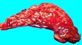
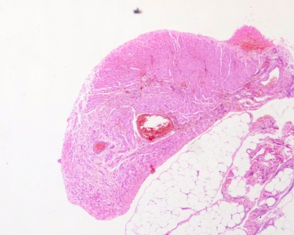
Segmental aplasia of the horn of a uterus. All that remains is a thin strip of tissue that is smooth muscle.

DSD can uterus hypoplasia

Histology of a bitch with uterine segmental aplasia. The only tissue present is smooth muscle attached to the mesometrium (YB157432).

McEntee K (1990) Reproductive Pathology of Domestic Mammals. Academic Press p 120-121

Mir F, Fontaine E, Albaric O, Greer M, Vannier F, Schlafer DH, Fontbonne A. Findings in uterine biopsies obtained by laparotomy from bitches with unexplained infertility or pregnancy loss: an observational study. Theriogenology. 2013; 79: 312-322.

McIntyre RL, Levy JK, Roberts JF, Reep RL (2010) Developmental uterine anomalies in cats and dogs undergoing elective ovariohysterectomy. J Amer Vet Medl Assoc 2010, 237 (5): 542-546.

Nakamura K, Yamasaki M, Osaki T, Ohta H, Sasaki N, Aoshima K, Kimura T, Takiguchi M. (2012) Bilateral Segmental Aplasia with Unilateral Uterine Horn Torsion in a Pomeranian Bitch. J Am Anim Hosp Assoc 2012, 48: 327-330

Oh K-S, Son CH, Kim B-S, Hwang S-S, Kim Y-J, Park Y-J, Jeong J-H, Jeong C, Park SH, Cho K-O. (2005) Segmental aplasia of uterine body in a adult mixed breed dog. J Vet Diag Invest. 17: 490-492, 2005

Ortega-Pacheco A, Segura-Correa JC, Jimenez-Coello M, Linde Forsberg C. (2006) Reproductive patterns and reproductive pathologies of stray bitches in the tropics. Theriogenology.

Rousset N, Abbondati e, Posch B, Owen LJ, Herrtage M (2011) Unilateral hydronephrosis and hydroureter secondary to ureteric atresia, and uterus unicornis in a young terrier. J Small Animal Pract 2011 52: 441–444.

Schulman ML, Bolton LA. (1997) Uterine horn aplasia with complications in two mixed-breed bitches. J S Afr Vet Assoc. 68(4): 150-153.


Testicular

Campos et al (2011) reported on a French bulldog with an XX genotype, that was SRY negative and which had testes. It was phenotypically male.

Silversides et al (2011) reported on a French bulldog with a female phenotype and an enlarged clitoris. The chromosomes were XX, the gonads testes and there was no SRY gene.

Buijtelsa et al (2013) reported on 10 dogs with DSD. 4 were XX, SRY- testicular DSD with uteri.

Del Carro et al (2014) reported on an 8 month old Miniature Pincher dog with scrotal testes, female but ambiguous external genitalia and enlarged clitorus. The internal genitalia was uterus and deferent duct.

Yoon et al (2018) reported on a young dog with an XX, SRY negative genotype, testes and female tubular genitalia. The tubular genitalia ended in the urethra, and externally the dog had ambiguous external genitalia and a large clitoris.

Schneider et al (2019) provided a detailed report of a case of an Australian Kelpie that was XX chromosomal type who had testes and was female in type with a large clitoris/hypoplastic penis.

Horňáková et al (2019) did a detailed analysis of a dog with cryptorchid testes, uterus and female appearance with enlarged clitoris. It was XX, SRY-, with uterus and enlarged clitoris.

Nowacka-Woszuk et al (2022) reported on a missense polymorphism in PADI6 in XX DSD SRY negative in French Bulldogs and American Staffordshire Terriers. They also looked at XY DSD Yorkshire Terriers but found no such polymorphism. PADI6 is peptidyl arginine deiminase 6 is a protein coding gene involved in cytoskeletal reorganization in the egg and early embryo development.


Buijtelsa JJCWM, de Giera J, Kooistraa HS, Grinwisb GCM, Naana E, Zijlstrac CC, Okkensa AC. (2012) Disorders of sexual development and associated changes in the pituitary-gonadal axis in dogs. Theriogenology 2013, 78: 1618–1626.

Campos M, Moreno-Manzano V, García-Roselló M, García-Roselló E(2011) SRY-Negative XX Sex Reversal in a French Bulldog. Reprod Dom Anim 2011 46: 185–188.

Del Carro AP, Rosset E, Josson-Schramme A, Lambert V, Buff S. First description of scrotal testicles in a dog affected by 78, XX testicular disorder of sex development. Reprod Dom Anim 2014; 49 c48-c52

Horňáková Ľ, Dianovský J, Holečková B, Šiviková K. A comprehensive study of disorder of sex development in Staffordshire bull terrier dog. Reprod Domest Anim. 2019; 54: 928-935.

Nowacka-Woszuk J, Stachowiak M, Szczerbal I, Szydlowski M, Szabelska-Beresewicz A, Zyprych-Walczak J, Krzeminska P, Nowak T, Lukomska A, Ligocka Z, Biezynski J, Dzimira S, Nizanski W, Switonski M. Whole genome sequencing identifies a missense polymorphism in PADI6 associated with testicular/ovotesticular XX disorder of sex development in dogs. Genomics. 2022; 114: 110389.

Schneider XJ, Fraser NS, Hill J. Theriogenology Question of the Month. J Amer Vet Med Assoc 2019; 254: 1401-1405

Silversides DW, Benoit JM, Collard F, Gilson C. Disorder of sex development (XX male, SRY negative) in a French bulldog. Can Vet J. 2011; 52: 670-672.

Yoon H, Han SH, Kim J, Kim K, Eom K. Urogenital anomalies and urinary incontinence in an English Cocker Spaniel dog with XX sex reversal. J Vet Intern Med 2018; 32: 1166-1171.

Ovotesticular

Bartel et al (2015) investigated receptors of estrogen, progesterone and testosterone in the tissues of a pug dog with XX ovotesticular DSD. The female tubular genitalia had progesterone receptor expression on the luminal epithelium but AR and ER in the glands and stroma of the endometrium. Sertoli cells, interstitial cells and stroma were AR positive. B catenin was positive in Sertoli cells but negative in normal dog Sertoli cells.

Bartel C, Meyer F, Schäfer-Somi S, Walter I. (2015) Expression of steroid hormone receptors in the genital structures of a true hermaphrodite pug dog. Reprod Domest Anim. 2015; 50: 164-167.

Other


XY disorders of sexual development

Gonadal dysgenesis

Ovarian

Testicular


Phenotype female

Herndon et al (2012) reported on a dog with an XY genotype, testes but a female phenotype that had testicular neoplasia – a Sertoli cell tumour in one gonad and a contralateral seminoma in a small testis. The dog had a female phenotype.


Phenotype male

General

Buijtelsa et al (2013) reported on 10 dogs with DSD. 3 were XY, SRY+ testicular DSD with uteri and os clitoris.


McGill J, Thieman Mankin KM, Parambeth JC, Edwards J, Cook A. Urine-Filled Large Prostatic Cystic Structure in Two Unrelated Male Miniature Dachshunds. J Am Anim Hosp Assoc. 2018; 54: e546-06


Cryptorchidism

Canine cryptorchidism is reviewed by Romagnoli (1991) and by Khan et al (2018).

Retained testes are seen sporadically as an isolated event, or they are seen in dogs with other tract anomalies, for example
miniature schnauzers with persistent Mullerian duct syndrome (PMDS) often have retained testes.

On rare occasions, the testis and epididymis are retained in an unusual location. Ectopic testes in dogs occur near the inguinal canal near the penis (Bloom 1954).

Those dogs with an increased risk, and for which the disease is familial include the boxer, cairn terrier, chihuahua, bulldog, maltese, mini dachshund, mini poodle, mini schnauzer, pekanese, pomeranium, Sheltand Sheep dog, toy poodle, and Yorkshire terrier. The heritibility appears to be autosomal recessive.

From a pathogenesis point of view (with the view to being able to provide hormonal therapy), the outgrowth and control of the gubernaculum and the cranial gonadal ligament are important. Romagnoli (1991) reviewed canine cryptorchidism and listed 3 different pathogenic mechanisms including absolute or relative failure of gubernacular outgrowth, aberrent growth with unusual position of gubernaculum, and excessive growth and absent or delayed regression of the gubernaculum. The exact influences (including hormonal influences) is largely unknown.

There is a tendency for most cryptorchids to be unilateral and right sided, with inguinal retention being more common than intraabdominal retention (James and Heywood 1979, Yates et al 1985). Ortega-Pacheco (2006) reported on 21 cases on 318 stray dogs and found 20 unilateral cases with an equal number on each side.

There are several complications with cryptorchidism including testicular neoplasia, especially Sertoli cell tumour and seminoma (Reif and Brodey 1969, Pendergrass and Hayes 1975), torsion of the testis, and complications of castration. These diseases are discussed under the relevant section on disease of the canine testis.

Cryptorchid testes are hypoplastic and as such are the same size as prepubertal testes, at least initially. They will become degenerate and therefore atrophy and become even smaller with time. The size of the epididymis is as expected for a prepubertal epididymis. It is assumed that atrophy of a retained testis occurs for the same reason that it becomes hypoplastic and spermatogenesis does not develop.


Figure : Retained testis with lack of spermatogenesis (hypoplasia).

Figure : Retained testis with lack of spermatogenesis (hypoplasia).

Bloom F (1954) Pathology of the dog and cat - The genitourinary system, with clinical considerations. American Veterinary Publications, Inc, Evanston Illinois. p215

Gharagozlou F, Youssefi R, Akbarinejad V, Mohammadkhani NI, Shahpoorzadeh T, Anti-Müllerian hormone: a potential biomarker for differential diagnosis of cryptorchidism in dogs. Vet Rec 2014; 175: 460

Hayes HM, Wilson GP, Pendergrass TW, Cox VS. (1985) Canine cryptorchidism and subsequent testticular neoplasia: case control study with epidemiologic update. Teratology 32: 51-56.

Kersten W, Molenaar GJ, Emmen JMA, Van der Schoot. (1996) Bilateral cryptorchidism in a dog with persistent cranial testis suspensory ligaments and inverted gubernacula: report of a case with implications for understanding normal and aberrant testis descent. J Anat 189: 171-176.

James RW, Heywood R. (1979) Age-related variations in the testes and prostate of beagle dogs. Toxicology. 12(3):273-279.

Marshall LS, Oehlert ML, Haskins ME, Selden JR, Patterson DF (1982). Persistent Mullerian duct syndrome in Miniature Schnauzers. J Amer Vet Med Assoc 181: 798-801

Memon M, Tibary A. Canine and feline cryptorchidism. In: Recent advances in small animal reproduction. PW Concannon, G England and J Verstegen (Eds) International Veterinary Information Service (www.ivis.org). Ithaca NY.

Ortega-Pacheco A, Rodríguez-Buenfil JC, Segura-Correa1 JC, Bolio-Gonzalez ME, Jiménez-Coello M and Linde Forsberg C. (2006) Pathological Conditions of the Reproductive Organs of Male Stray Dogs in the Tropics: Prevalence, Risk Factors, Morphological Findings and Testosterone Concentrations. Reprod Dom Anim 41 :429-437

Pendergrass TW, Hayes HM ((1975) Cryptorchidism and related defects in dogs: epidemiologic comparisons with man. Teratology 12: 51-56.

Reif JS, Brodey RS (1969) The relationship between cryptorchidism and canine testicular neoplasia. J Amer Vet Med Assoc 155: 2005-2010.

Romagnoli SE. (1991) Canine cryptorchidism. Vet Clinics of North Amer: Small Animal Pract. 21: 533-544.

Yates D, Hayes G, Heffernan M, Beynon R. 2003 Incidence of cryptorchidism in dogs and cats. Vet Rec 152(16):502-504.

Hypospadias

Hypospadia is the complete or partial failure of fusion of the frenulum of the penis and or prepuce. At its extreme, there is a bifid scrotum. Severe cases can also have fusion of the urethra and rectum.

Waller et al 2021 reported a case with extensive failure of fusion , a bifid scrotum, perineal fissure and poorly formed penis and a prepuse not fused to the abdominal wall.


Mestrinho LA, Alberto A, Iglésias LV, Gordo I. Cloacal malformation in a 5-month-old male dog. Can Vet J. 2019 Dec;60(12):1291-1294.

Waller SB, Neto LHSB, Costa PPC, Teles CHA, De Alencar MP, Freitas LA, Cleff MB, De Araújo AA. Hypospadias in a mongrel male (78,XY; SRY-positive) dog. J Small Anim Pract. 2021; 62: 65.

Persistent Mullerian duct syndrome

Miniature Schnauzers are recognised to have a common syndrome where the male dogs develop uteri. They have an XY genotype (although some may be XXY), testes and a male phenotype but have persistence of the paramesonephric duct system. They are deficient in the AMH - receptor interaction. Uni or bilateral cryptorchidism and Sertoli cell tumours and endometritis are part of the syndrome.

In miniature Schnauzers, the defect is in the AMHR2 gene. It is a C to T transition (C241T) in exon 3 of the Müllerian inhibiting substance type II receptor gene (aka AMHR2). The prevalence in miniature Schnauzers is such that the AMHR2 mutation allele frequency is reported to be 0.16 and a carrier genotypic frequency is 0.27


Myerscough (1993) reports on a German Shepherd dog that was male, but which had pyometra and torsion of the uterus.

Breshears and Peters (2011) wrote a case report about a miniature schnauzer with cryptorchidism and a uterus.

Kim YJ, Kwon HJ, Byun HS, Yeom D, Choi JH, Kim JH, Shim H. Surveyor assay to diagnose persistent Müllerian duct syndrome in miniature Schnauzers. J Vet Sci. 2016

Pujar S, Meyers-Wallen VN. A molecular diagnostic test for persistent Müllerian duct syndrome in miniature schnauzer dogs. Sex Dev. 2009;3(6):326-8.

Smit MM, Ekenstedt KJ, Minor KM, Lim CK, Leegwater P, Furrow E. Prevalence of the AMHR2 mutation in Miniature Schnauzers and genetic investigation of a Belgian Malinois with persistent Müllerian duct syndrome. Reprod Domest Anim. 2018; 53: 371-376.

Prostatic utricle

McGill et al (2018) reported on 2 unrelated miniature Dachshunds with cysts dorsal to and which communicated with the urethra. They were considered prostatic utricles. These dogs also had 'dysplastic testicles' and the other had 'testicular hypoplasia and epididymal fibrosis without spermatogenesis'.


McGill J, Thieman Mankin KM, Parambeth JC, Edwards J, Cook A. Urine-Filled Large Prostatic Cystic Structure in Two Unrelated Male Miniature Dachshunds. J Am Anim Hosp Assoc. 2018; 54: e546-06


Testicular hypoplasia

Testicular hypoplasia is where the testis does not develop to its normal size. It is always accompanied by a failure of the epididymis to acquire its normal size. Most cases of hypoplasia are because of cryptorchidism, but in this section, primary hypoplasia is limited to those situations where the testis has descended normally. Hypoplasia is almost always seen as a failure of the prepubertal testis to enlarge, but there may be cases where even the prepubertal testis is smaller than normal. Hypoplasia is best diagnosed clinically by identifying that the testis has not increased in size from puberty, but this change is seldom monitored in dogs and so the presence of a small testis could mean either hypoplasia or atrophy. Atrophy is an acquired condition, but hypoplasia is potentially a genetic and heritable condition.

Hypoplasia may accompany chromosomal abnormalities. Nie et al (1998) reported a case of canine Klinefelters syndrome (XXY) that had testicular hypoplasia. Others are not. Metcalfe et al (1999) reported on 2 Labrador retrievers that had testicular hypoplasia. Their testes were half the weight of a control dog. Both dogs had lymphocytic intersitial orchitis. l. Ortega-Pacheco et al (2006) found 21 cases in 318 stray dogs. 14 were bilateral, 1 was right unilateral and 6 were left unilateral in distribution.

Hypoplasia is variable in its degree. In production animals, testicular volume or scrotal circumference is measured and a certain size is deemed to be the minimum acceptible. Smaller testes than the minimum size have some degree of hypoplasia - in some, there are only mild changes, but in others, they are extreme.

The histological appearance of hypoplasia is also variable. Affected animals have seminiferous tubules that lack complete spermatogenesis. In the most hypoplastic testes, all the tubules lack germ cells or any spermatogenesis. Other individuals have some stages of spermatogenesis but spermatogenesis stops at a particular stage such as at the primary spermatocyte stage. These are said to have spermatogenic arrest. Other individuals have some normal tubules and some tubules that are arrested. Where there is arrested development, the cells may all undergo apoptosis. Unfortunately, testicular degeneration has an identical change, so other factors must be used to determine if the change is due to hypoplasia or not.

Most references to hypoplasia do so in the context of azoospermia (see section on azoospermia above). Rehm (2000) found 30% of control beagle dogs to have segmental hypospermatogenesis, and 18% of dogs to have tubules with 'degeneration/hypoplasia", so clinically normal dogs can have undetected changes. The general lack of detailed studies on testicular hypoplasia in dogs indicates this to be a sporadic condition when it is the only anomaly.


Metcalfe SS, Gunn IM, Chamness KA. (1999) Azoospermia in two Labrador Retrievers. Aust vet J 77: 570-573.

Nie GJ, Johnston SD, Hayden DW, Buoen LC, Stephens M (1998) Theriogenology question of the Month. J Amer Vet Med Assoc 212: 1545-1547.

Ortega-Pacheco A, Rodríguez-Buenfil JC, Segura-Correa1 JC, Bolio-Gonzalez ME, Jiménez-Coello M and Linde Forsberg C. (2006) Pathological Conditions of the Reproductive Organs of Male Stray Dogs in the Tropics: Prevalence, Risk Factors, Morphological Findings and Testosterone Concentrations. Reprod Dom Anim 41 :429-437

Rehm S. (2000) Spontaneous testicular lesions in purpose-bred beagle dogs. Toxicol Pathol. 28(6):782-787.


Hypospadias


JAVMA 227: 887-888
ACT Theriogenology question of month
Adult Papillon


Can Vet J 46: 1022-1025
Hypospadias in a 1 yr old WHWT
Perineal opening.


References


Breshears MA, Peters JL. (2011) Ambiguous Genitalia in a Fertile, Unilaterally Cryptorchid Male Miniature Schnauzer Dog. Vet Pathol 2011, 48: 1038-1040.

Campos M, Moreno-Manzano V, García-Roselló M, García-Roselló E(2011) SRY-Negative XX Sex Reversal in a French Bulldog. Reprod Dom Anim 2011 46: 185–188.

Del Carro AP, Rosset E, Josson-Schramme A, Lambert V, Buff S. First description of scrotal testicles in a dog affected by 78, XX testicular disorder of sex development. Reprod Dom Anim 2014; 49 c48-c52

Gharagozlou F, Youssefi R, Akbarinejad V, Mohammadkhani NI, Shahpoorzadeh T, Anti-Müllerian hormone: a potential biomarker for differential diagnosis of cryptorchidism in dogs. Vet Rec 175: 460 

Herndon AM, DVM, Casal ML, Jaques JT (Scott) (2012) Testicular Neoplasia in the Retained Testicles of an Intersex Male Dog. J Am Anim Hosp Assoc 2012; 48:118–124.

Melniczek JR, Dambach D, Prociuk U, Jezyk PF, Henthorn PS, Patterson DF, Giger U. Sry-negative XX sex reversal in a family of Norwegian Elkhounds.J Vet Intern Med. 1999 Nov-Dec;13(6):564-9. Review.

Meyers-Wallen VN, Donahoe PK, Ueno S, Manganaro TF, Patterson DF. (1989) Mullerian inhibiting substance is present in testes of dogs with persistent Mullerian dict syndrome. Biol of Reproduct 41: 881-888.

Meyers-Wallen VN, Patterson DF (19) Disorders of sexual development in dogs and cats. Vet Clinics of North Amer:Small Anim Pract 21: 1260-1269.

Meyers-Wallen VN (2001) Inherited abnormalities of sexual development in dogs and cats. In: Recent advances in small animal reproduction. PW Concannon, G England and J Verstegen (Eds) International Veterinary Information Service (www.ivis.org). Ithaca NY.

Meyers-Wallen VN (2009) Review and update: genomic and molecular advances in sex determination and differentiation in small animals. Reprod Dom Anim 44: 40-46

Marshall LS, Oehlert ML, Haskins ME, Selden JR, Patterson DF (1982). Persistent Mullerian duct syndrome in Miniature Schnauzers. J Amer Vet Med Assoc 181: 798-801

Myerscough M (1993) Pyometra and torsion of the uterus in a male dog. Vet Rec 133: 252

O'Connor CL, Schweizer C, Gradil C, Schlafer D, Lopate C, Prociuk U, Meyers-Wallen VN, Casal ML. (2011) Trisomy-X with estrous cycle anomalies in two female dogs. Theriogenology. 2011, 76: 374-380.

Rota A, Starvaggi Cucuzza A, Iussich S, DeLorenzi L, Parma P (2010) The Case of an Sry-Negative XX Male Pug With an Inguinal Gonad. Reprod Dom Anim 2010 45:743-745

Sommer MM Meyers-Wallen VN. (1991) XX true hermaphroditeism in a dog.J Amer Vet Med Assoc 198: 435-438.

Williams J, Partington BP, Smith B, Law JM (1997) pyovagina and stump pyometra in a neutered xx sex-reversed Beagle: a case report. J Amer Anim Hosp Assoc 33: 83-90.