This page is part of the site called Surgical Pathology of the Canine Male Reproductive Tract by

Dr Rob Foster
OVC Pathobiology
University of Guelph

© All materials are copyright and should only be used with the expressed permission of the author.


Disease of the Spermatic Cord


The spermatic cord is a structure composed of the testicular artery and veins that form the pampiniform plexus, lymphatics and the deferent duct. It is covered by the vaginal tunic and by the cremaster muscle. Disease of this area is rare, or accompanies disease of the adjacent tissues.

Degenerative vascular changes

Torsion of the spermatic cord

When there is a twisting of the testis on its axis, the torsion is of the spermatic cord. This is commonly called testicular torsion, and will be dealt with under testicular torsion

Varicocele

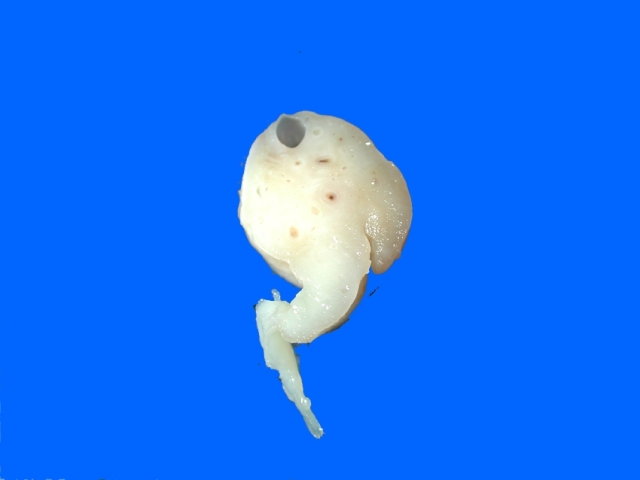
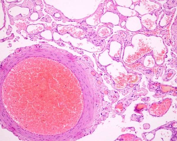
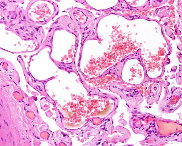
A varix or varices of this area is called a varicocele. It can be a small structure comprised of dilated veins, but usually those that are clinicaly identified are large and thrombosed. Varicocele is common in humans as the left testicular vein terminates at the left renal vein, and the insertion site causes increased pressure in this location. Spontaneous cases of primary varicocele in dogs are not reported, but dogs are used as an experimental model for varicocele in man. Increasing pressure in the spermatic vein has been done by surgical ligation.

There are two cases in the YB database (YB20491, 45435) where histologically there were dilated thrombosed veins in the pampiniform plexus. Examination of the formalin fixed tissues of one case confirmed this as a varicocele. There was necrosis and fibrosis in the tissues suggesting trauma. No other evidence of torsion was seen, so injury to the vessels, fibrosis and an increased venous pressure was thought to be the pathogenesis. The cause of the second case is not known.

Secondary varicocele formation is reported. McNeil and Weaver (1980) reports varicosities developing secondary to metastatis of Sertoli cell tumors.


McNeil PE, Weaver AD. (1980) Massive scrotal swelling in two unusual cases of canine Sertoli cell tumour. Vet Rec 106: 144-146.

Turner TT. (2001) The study of varicocele through the use of animal models. Hum Reprod Update. 7(1):78-84.

Ushio N, Chambers JK, Watanabe K, Kayano M, Uchida K. Age-Related Arteriolar Changes With Lipid and Amyloid Deposits in the Gonads of Dogs. Vet Pathol. 2021; 58: 558-567.

Atherosclerosis

Atherosclerosis is unusual in dogs, and is seen in hypothyroidism and in diabetes mellitus. While these causes of atherosclerosis are well recongised, the testicular arteries and veins and seldom examined. No cases were found in the PubMed database. Cases no doubt occur and will eventually be seen.

Ushio et al (2021) reported atherosclerosis (they classified it as vacuolar change) in 2 of 72 dogs

Ushio N, Chambers JK, Watanabe K, Kayano M, Uchida K. Age-Related Arteriolar Changes With Lipid and Amyloid Deposits in the Gonads of Dogs. Vet Pathol. 2021; 58: 558-567.

Fibromuscular hypertrophy

Ushio et al (2021) reported fibromuscular hypertrophy in 13 of 72 dogs

Ushio N, Chambers JK, Watanabe K, Kayano M, Uchida K. Age-Related Arteriolar Changes With Lipid and Amyloid Deposits in the Gonads of Dogs. Vet Pathol. 2021; 58: 558-567.

Hyalinosis

Ushio et al (2021) reported hyalinosis in 5 of 72 dogs

Ushio N, Chambers JK, Watanabe K, Kayano M, Uchida K. Age-Related Arteriolar Changes With Lipid and Amyloid Deposits in the Gonads of Dogs. Vet Pathol. 2021; 58: 558-567.

Vasculitis

Vasculitis was only seen in the epididymis, but the exact location of the vessels were not given. it was seen in 10 of 72 dogs

Ushio N, Chambers JK, Watanabe K, Kayano M, Uchida K. Age-Related Arteriolar Changes With Lipid and Amyloid Deposits in the Gonads of Dogs. Vet Pathol. 2021; 58: 558-567.

Inguinal hernia

This topic is covered under scrotum and tunics

Inflammation

Inflammation of the spermatic cord is called funisitis or funiculitis. Inflammation of the cord is unusual. It has been reported in Brucella canis infection (Brennan et al 2008). Complications of castration with contamination of the spermatic cord with bacteria or foreign material is unusual with the appropriate surgical procedures.
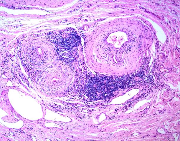
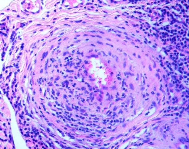
Vasculitis

Vascultitis of the vessels is reported and seen occasionally. It is reported in beagles with systemic vascilitis (canine juvenile polyarteritis syndrome (beagle pain syndrome).) (Albassam et al 1989) and periodically in other animals. Primary signs are unusual, but ischemic changes in the testis incuding degeneration occur. No gross lesions are reported to the spermatic cord, but histological changes can be dramatic.

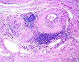
Figure : Vasculitis of the testicular artery (YB144756)

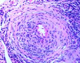
Figure : Arteritis of testicular artery

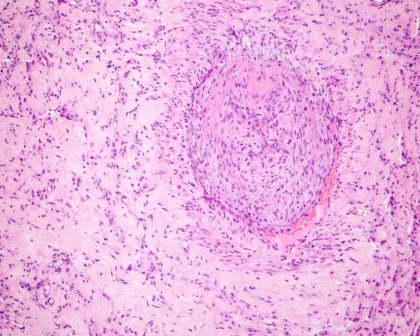
Fibrosis of the spermatic cord was an unusual finding in one dog (YB116854). This lesion is likely to be the end result of vasculitis.

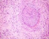
Figure :Fibrosis of spermatic cord and vascular hypertrophy/fibrosis (YB116854)


Albassam MA, Houston BJ, Greaves P, Barsoum N. (1989) Polyarteritis in a beagle. J Am Vet Med Assoc. 194(11):1595-1597.

Brennan SJ, Ngeleka M, Philibert HM, Forbes LB, and Allen AL (2008) Canine brucellosis in a Saskatchewan kennel. Can Vet J. 49(7): 703-708.

Neoplasia

Testicular tumours

Extratesticular testicular tumors have been found in this locaton (see Testicular neoplasms). They presumably arose in the castrated dogs fromcontinued stimulation of testicular remnants by pituitary hormones lacking negative feedback. In dogs, most of the testicular neoplasms in the spermatic cord were Sertoli cell tumours.

Metastatic testicular tumours, although rare, may be found in the spermatic cord and it is the finding of metastases in this location that confirms the tumour as being metastatic. Sertoli cell tumors expecially, but also seminoma and interstitial cell tumours may metastasize to this location.

Primary tumors of the spermatic cord

Primary neoplasms of any of the tissue in the spermatic cord could arise from the spermatic cord, or metastasize there from any malignant neoplasm. Here are some examples.

Epithelial Neoplasms


Carcinoma

Some of the most widely metastatic neoplasms of dogs are transitional cell carcinomas, and as such, metastasis to any tissue is possible. One metastatic carcinoma to the spermatic cord and vaginal tunic is in the YB database (YB162821). It is described above (see tunics) as it was manifested by carcinomatosis of the tunics.


Figure : Pampiniform plexus of a dog infiltrated with a scirrhous carcinoma.

Mesenchymal

Haemangioma

With the large amount of vascularity in the spermatic cord, neoplasia of vascular origin is virtually a given! There is one case in the YB database (YB147752) where a dog had a proliferation of vessels within the spermatic cord. This was identified as a thickening of the spermatic cord, and grossly as a red to black area of swelling and discolouration. These did not form a mass, but infiltrated the cord. They had the same appearance as a haemangioma.


Sarcomas

McEntee (1990) reports seeing 'malignant neoplasms of adipose tisue, fibrous tissue and striated muscle'. He indicated finding metastases in the regional lymph nodes.

McEntee (1990). Reproductive Pathology of Domestic Mammals. Academic Press. p281.

Round cell neoplasms

Lymphoma

Brunati et al (2023) reported on a dog with a mass of the spermatic cord that was cytologically and histologically identified as a lymphoma. It was a B cell lymphoma. At later time, it also involved the brain.

Brunati G, Pintore L, Avallone G, Tomba A, Cantile C, Valenti P. A case of spermatic cord B-cell lymphoma relapsing to the brain in a dog. Canadian Vet J 2023; 64: 529-533.

Miscellaneous

Ectopic spleen

There is one case in the YagerBest Database where ectopic spleen was seen in the pampiniform. The small nodule was identical to ectopic spleens seen in the mesentry.