This page is part of the site called Surgical
Pathology of the Canine Male Reproductive Tract
by
Dr Rob Foster
OVC Pathobiology
University of Guelph
Table of Contents
General Considerations
Surgical pathology is a field that combines the traditional discipline of
pathology with a keen sense of the clinical picture and prognosis. It is generally
focused on the individual animal and thus concentrates on companion animals
- mostly dogs and cats. This 'chapter' focuses on materials that would be
sent by a clinician to laboratory specializing in surgical biopsies. An accurate
diagnosis is fundamental to prognosis, so this section concentrates on reaching
an accurate diagnosis so that the prognosis will become obvious.
Diagnoses are best made with a clear understanding of normal functional anatomy,
so a section on normal anatomy is included.
The link is above.
There are many conditions and diseases that are of a congenital nature, and
while many of these are of little consequence, some are important. There is
a page dedicated to Disorders of Sexual Development and this can be accessed from the link on the left.
Many male dogs are castrated before they reach puberty. As a result, very
few lesions are to be found in what remains of the male reproductive tract
- carcinoma of the prostate is one exception.
The vast majority of the cases illustrated here are those seen through the
surgical biopsy service of Yager-Best
Veterinary Surgical Pathology, Guelph, Ontario, (aka YagerBest Histovet) and I am indebted
to Drs Julie Yager and Susan Best for the use of this material. The designation
YB and number is the case number of the particular sample.
There are 2 excellent text books
that contain a wealth of information. Bloom (1954) and McEntee (1990) are
2 most comprehensive texts.
Bloom F (1954) Pathology of the
dog and cat - The genitourinary system, with clinical considerations. American
Veterinary Publications, Inc, Evanston Illinois.
McEntee K (1990) Reproductive
Pathology of Domestic Mammals. Academic Press.
Sample
selection and fixation
Surgical pathologists
are asked to examine materials from the reproductive tract or where the
components of the reproductive tract once were. The sample is usually provided
as a surgical specimen and is small enough to warrant no further processing
than placing in a small jar of formalin ensuring the volume of fluid to
sample is at least 10 to 1.
Examination of tissues
by submitting veterinarians is encouraged but butchery of tissues is not.
The whole testes and epididymides will not usually fit into a standard surgical
biopsy jar, so some sectioning is necessary. Here is an example of what
not to do!
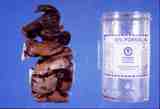
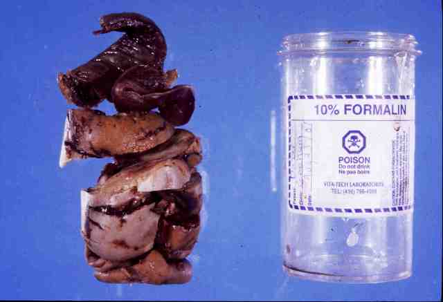
Figure 1: Biopsy submission of testis and epididymis
from a dog. Testis is cut in many pieces and would just fit in the jar.
There was room for a couple drops of formalin!
The best way to sample the testis and epididymis is to cut the sample sagitally.
For the majority of cases, this can be placed in a container of formalin
with 10 parts liquid to one part tissue. If necessary, a smaller sample
can be collected for examination, or all parts can be sent for the pathologist
to examine.
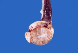
Figure 2: Recommended method for cutting testis for examination.
It is common practice
for technicians to trim tissues for histopathology. Submission of all of
the tissue will allow the pathologist to examine it if necessary. In those
cases were the clinical history does not match the histological findings,
examination of the fixed tissue is an essential component of arriving at
the correct diagnosis.
Complications
of castration surgery
Castration
is such a common procedure and the scrotal contents so efficiently packaged
that it complications are rare and surgical specimens are rarely taken.
It is worthwhile though listing some of the complications so that there
is a list of differential diagnoses (diagnostic hypotheses) to consider
should such a specimen be received. This is a short list of complications
of which I am aware.
- adhesions
- anaesthesia
- arterovenous anastosmosis.
- dehiscence
- extratesticular testicular tumours
- foreign body reaction to entrapped hair.
- haemorrhage
- inadvertant prostatectomy
- infection
- pain
- scrotal oedema, haemorrhage
-
testicular remnants
Most of this list is self explanatory.
Aiken et al (1993) reports on a dog that developed an arteriovenous fistula
of the spermatic cord after castration. The lesion was a 3 x 10cm pulsatile
mass. Surgical excision was curative.
Extratesticular testicular tumours will be dealt with below. Briefly, previously castrated
dogs have developed Sertoli cell tumours in many cases, many years after
being castrated. This is very rare.
The removal of inguinally or abdominally retained gonads is much more problematic,
and so are the complications. One reported complication is inadvertant removal
of the prostate, where the deferent duct (ductus deferens) is followed to
the end, and the prostate is removed! This is reported by Schultz et al
(1996) and Vititoe et al (2013) and was seen in a case in 2005 (Riley survived).
Testicular remnants will
be discussed below in the section on "Disease within the abdomen".
Hamilton at al (2014) compared and closed orchidectomy and the complications that occur with each. The complications they report include "incisional haemorrhage,
incision discharge, incision swelling, incision bruising, incision, erythema, incision dehiscence, incision pain, scrotal bruising, scrotal swelling, scrotal pain, pyoderma, diarrhoea, and vomiting."
Aiken SW,
Jakovljevic S, Lantz GC, Blevins WE. (1993) Acquired arteriovenous fistula
secondary to castration in a dog. J Am Vet Med Assoc.202:965-7.
JAVMA 208 (11) 1882
Hamilton KH, Henderson ER, Toscano M, Chanoit GP. (2014) Comparison of postoperative complications in healthy dogs undergoing open and closed orchidectomy. J Small Anim Pract 2014; 55(10): 521-526.
Schultz KS, Waldron
DR, Smith MM, Henderson RA, Howe LM. (1996). Inadvertant prostatectomy as
a complication of cryptorchidism in four dogs. J Amer Anim Hosp Assoc 32:
211-214.
Vititoe K, et al. (2013) Anuria due to inadvertent prostatectomy during cryptorchidectomy. Can Vet J 2013;54:675-677.