Normal Reproductive Tract of the Queen/female Cat
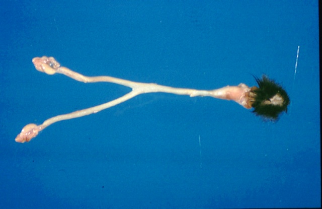
Figure : Normal reproductive tract
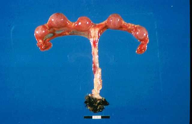
Figure :Normal Reproductive tract, pregnancy
Figure : Normal reproductive tract
Figure :Normal Reproductive tract, pregnancy
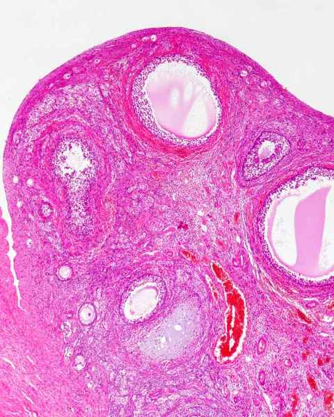
The normal ovary of the queen is a simple structure and there are few unusual structures, apart from the interstitial cells of the ovary. The surface of the ovary is smooth and the follicles and corpus lutea cause only slight undulaton of the surface. Multiple follicles develop and although they are induced ovulators, it is common to see corpora lutea in animals that were not bred. So called noncopulatory ovulation is apparently common (Gudermuth et al 1997)
The ovary is suspended from the dorsal abdominal wall by the mesovarium within which is a pampiniform plexus of blood vessels and which has an outer covering of smooth muscle. This latter structure is present in the mesometrium and is usefull for tissue identification when only small pieces of tissue are presented for biopsy.
Figure : Normal feline ovary
Figure :Normal adult feline ovary
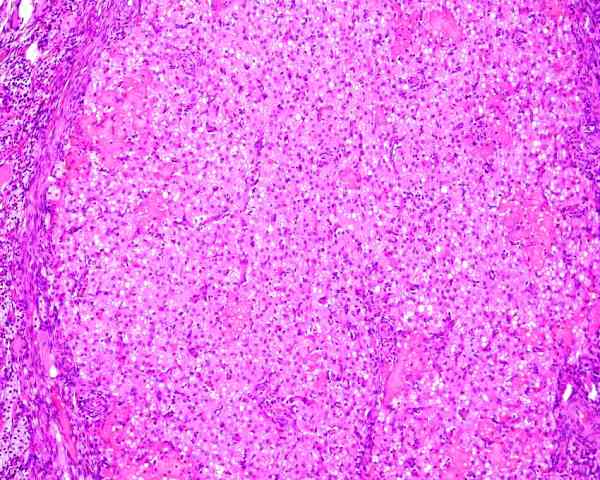
Figure : Normal corpus luteum
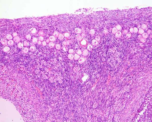
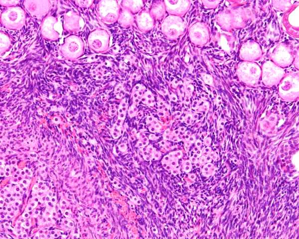
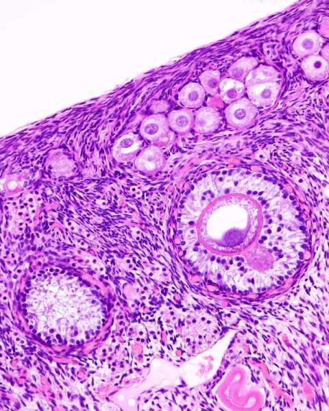
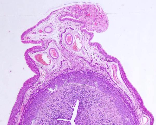
The ovary of juvenile cats is a little different to most other species. The germ cells (oocytes) cluster in a layer beneath the surface (or epithelium) of the ovary. With age, these reduce in number. The germ cells are surrounded by squamous follicular cells that presumably arise from the mesonephric tubules that grow into the ovary. Follicular cells are on a basement membrane. Together they form the primordial follicle. The follicular cells enlarge and become cuboidal - this is the primary follicle. These cuboidal follicular cells begin to proliferate, and become the granulosa cells. Beneath the basement membrane, stroma cells form a layer of cells called the follicular thecal layer, that subsequently proliferates and separates into the the internal and external thecal layers. When a follicle ovulates, the granulosa cells and the theca cells form populations of cells that form the corpus luteum (yellow body).
Another unique feature of the feline ovary is the presence of interstitial (endocrine) cells of the ovary. These are small uniform cells that form clusters or surround a degenerate follicle. The interstitial cells of young (immature cats) appear to develop from the ovarian medula. They also appear to arise from the theca interna of atretic follicles (Perez et al 1999)
Figure : Normal juvenile feline ovary. Germ cells form a layer beneath the surface. Interstitial cells present in the stroma.
Figure : Normal juvenile feline ovary. Clusters of interstitial cells are visible beneath the germ cells.
Figure : Normal juvenile feline ovary. Germ cells, primary follicle (right center) and interstitial cells are present.
It is common to see tubules lined by ciliated columnar or cuboidal cells. These are remnants of mesonephric tubules and form the rete ovarii. There are 3 parts to the rete ovarii - the intraovarian rete, the extraovarian rete, and a connecting rete. The rete are supposed to have a secretory activity. The intraovarian rete is supposed to be noncilited, whereas the connecting rete is lined by ciliated epithelial cells.
Figure :Hilus of the ovary with rete ovarii (mesonephric tubules)
Reynaud K, Gicquel C, Thoumire S, Chebrout M, Ficheux C,Bestandji M, Chastant-Maillard S (2009) Folliculogenesis and Morphometry of Oocyte and Follicle Growth in the Feline Ovary. Reprod Dom Anim 44: 174-17
Gudermuth DF, Newton L, Daels P, Concannon P. J (1997) Incidence of spontaneous ovulation in young, group-housed cats based on serum and faecal concentrations of progesterone. Reprod Fertil Suppl. 51:177-184.
Perez JF, Conley AJ, Dieter JA, Sanz-Ortega J, Lasley BL (1999) Studies on the origin of ovarian interstitial tissue and the incidence of endometrial hyperplasia in domestic and feral cats. Gen Comp Endocrinol. 116(1):10-20.
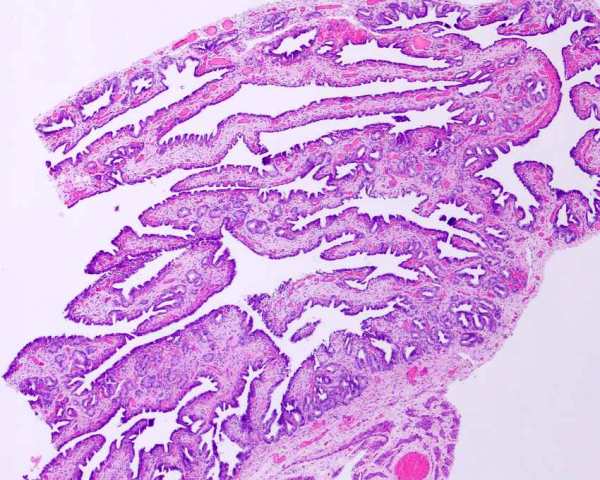
The uterine tube of the cat has several parts. The infundibulum or funnel of the uterine tube is over the surface of the ovary but does not surround the ovary as in the dog. There is an abdominal opening around which is a fringe of processes (fimbriae). The ampulla is the relatively wide part from the abdominal opening to the narrow portion of the tube (called the isthmus) There is a uterine part of the tube, and a uterine oriface. For the surgical pathologist, these have little relevance to diagnosis or distribution of lesions. The fringe of processes may be mistaken for neoplasia or hyperplasia, but the well differentiated appearance makes this unlikely. The uterine tube is attached to the body by a mesosalpynx.
Figure : Fringe of processes of the uterine tube/
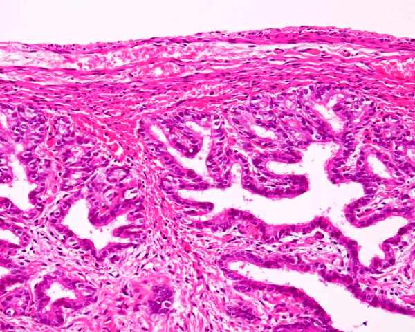
Figure : Fringe of processes (fimbriae) of the uterine tube.
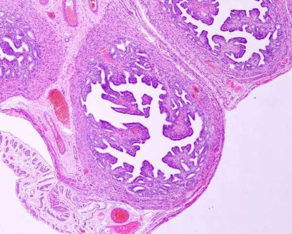
Figure : Uterine tube, narrow portion (isthmus).
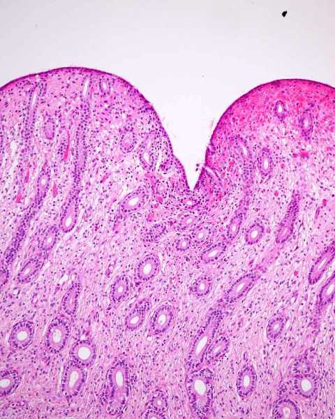
The normal uterus of the cat has 2 uterine horns, a uterine body and cervix. These are suspended into the abdominal cavity by the mesometrium. The surface of the feline uterus is usually smooth and regular (unlike the dog). When pregnant, bulges occur at regular intervals.
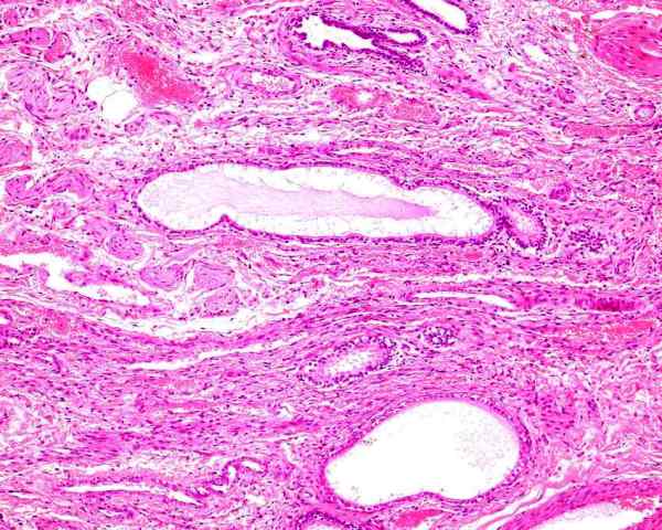
The endometrium is the inner portion of the uterus and it has luminal epithelium, glands and stroma. The myometrium is composed of an inner circular and oblique layer, a vascular layer, and an outer longitudinal layer that continues up the mesometrium beneath the serosa.
Chatdarong et al (2005) describes the histology of the normal endometrium of cats in various stages of the estrus cycle. There is a variation in height of the endometrium giving it an undulating profile in a sagital plain.
Figure : Feline uterus with endometrium, myometrium and serosa.
Figure : Feline mesometrium with blood vessels, serosa and a band of smooth muscle that is continuous with the longitudinal muscular layer of the myometrium.
Figure : Feline endometrium.
Chatdarong K, Rungsipipat A, Axner E, Linde Forsberg C. (2005) Hysterographic appearance and uterine histology at different stages of the reproductive cycle and after progestagen treatment in the domestic cat. Theriogenology. 64(1):12-29.
Clemetson and Ward (1990) reported their results of culturing of the vagina and uterus of healthy cats. They examined 13 multiparous adult cats, 10 kittens 5-7 months old, and 30 cats submitted for ovariohysterectomy. Aerobic bacteria were isolated from the vaginas of 52 of 53 cats and from 2 of 29 uterine swabs. Streptococci, Staphylococci, E. coli are the most common isolates.
Clemetson LL, Ward ACS (1990) Bacterial flora of the vagina and uterus of healthy cats. J Amer Vet Med Assoc 196: 902-906.
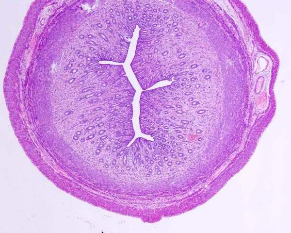
The cervix is actually part of the uterus - it is called the neck of the uterus. It has an internal orifice, central canal, and external oriface that is on the vaginal end where the cervix extends a short distance into the vagina. The glands of the cervix are not well developed and are mostly straight and short. The lining cells are identical to the luminal epithelium of the endometrium. The portion that projects into the vagina is lined on its outer surface by vaginal epithelium and by endometrium on its inner surface.
Zambelli D, Cunto M. (2005) Vaginal and cervical modifications during the estrus cycle in the domestic cat. Theriogenology. 64(3):679-684.
Zambelli and Cunto (2005) reviewed the anatomy of the vagina and cervix of the cat. The cat has a structure called a urogenital sinus with an entrance to the vagina and a urethral opening. Much of this is the vestibule and is a 4 mm diameter (ID) structure that is 1 to 2 cm long. The vagina is non distensible and about 1mm diameter (ID). There is a fornix ventral to the cervix. The vagina is about 2-3 cm long, so that the distance from the vulva to the cervix is about 4-4.5 cm long. The vagina is lined by stratified nonkeratinizing epithelium.
There are glandular acini within the wall of the lower part of the vagina (Shehata 1973). The acini are mucous and serous in type. The former were a tall columnar epithelium with flattened basal nuclei, and the latter were low columnar cells with basal nuclei. Some were within the submucosa and others were intermuscular. Unfortunately, the paper described the glands as being in the vagina but it was not clear if this was the actual vagina or part of the vestibule. The comment was made that the glands were analagous to Bartholin's glands of humans. I have histologically examined the vestibule and vulva of a cat and there are glands identical to apocrine glands in the deep tissues of the vulva that were separate to those of the vulval skin.
Shehata R (1973) Glandular acini in the wall of the lower part of the domestic cat vagina. Acta anat 84: 52-61.
Zambelli D, Cunto M. (2005) Vaginal and cervical modifications during the estrus cycle in the domestic cat. Theriogenology. 64(3):679-684.
Clemetson and Ward (1990) reported their results of culturing of the vagina and uterus of healthy cats. They examined 13 multiparous adult cats, 10 kittens 5-7 months old, and 30 cats submitted for ovariohysterectomy. Aerobic bacteria were isolated from the vaginas of 52 of 53 cats and from 2 of 29 uterine swabs. Streptococci, Staphylococci, E. coli are the most common isolates.
Clemetson LL, Ward ACS (1990) Bacterial flora of the vagina and uterus of healthy cats. J Amer Vet Med Assoc 196: 902-906.
The vulva of the cat has stratified nonsquamous epithelium on its inner surface and haired skin on the outer surface.