This page is part of the site called Surgical Pathology of the Feline Female Reproductive Tract by

Dr Rob Foster
OVC Pathobiology
University of Guelph

Disease of the Feline Vagina and Vulva

Table of Contents


General considerations

Lesions of the feline vagina and vestibule are rare. Those of the external skin of the vulva are a little more common, but not by very much! The small size of the external genitalia of the cat no doubt accounts for some of this. The fact that most pet cats are neutered early in life will account for much as well.

There are very few case reports of reproductive disease in the female cat, and anomalies account for most of them. The submissions to Yager Best Surgical Pathology include the following

Disease of the vagina and vestibule

Vaginitis
1
Neoplasia

carcinoma

1
leiomyoma
1
Polyp
1

Disease of the vulva

Neoplasia
apocrine carcinoma
6
anaplastic sarcoma
2
mast cell tumour
1

Inflammation

Inflammation of the vagina and vestibule should be the most common condition, as vaginitis and vulvitis would be expected to occur in endometritis and pyometra. Samples for histology of the vagina and vulva are not usually taken in these conditions, and the size of these structures are not conducive to routine biopsy evaluation.

Nicastro and Walshaw (2007) report on a cat with chronic vaginal discharge secondary to retention of the bones of a fetal kitten.

Nicastro A, Walshaw R (2007) Chronic Vaginitis Associated With Vaginal Foreign Bodies in a Cat J Am Anim Hosp Assoc 43: 352-355


Neoplasia of the vagina, vestibule and vulva

Neoplasia is very rare. Despite this, neoplasia was the most common lesion of the vulva found in a surgical pathology practice (YagerBest Histovet). Apocrine adenocarcinoma of the vulva was the most common at 7 cases. Followup information was available from 5 cases and all but one was euthanised 2-18 months after diagnosis for reoccurrence of local disease (3 cats) or signs attributed to metastasis (3 cases) (Welsh et al 2010). Two anaplastic sarcomas of the vulva were also seen, and 1 mast cell tumour is recorded.

In the vagina and vestibule, one carcinoma and one leiomyoma were found. None of these had special features.

Patnaik (1993) reports a single case of a granular cell tumour in the vulva of a cat.

Firat et al (2007) report a single case of a vulvar leiomyosarcoma. Surgical removal was attempted, but it regrew and the cat was later euthanised.

Conrado et al (2023) reported a case of adenocarcinoma of the left vulval fold. It had tissue eosinophilia with the neoplasms. Indications were of metastasis to a lymph node.


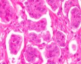
Figure : Apocrine adenocarcinoma of vulva in a cat.

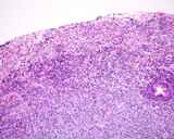
Figure : Anaplastic sarcoma of the vagina of a cat. The surface was ulcerated and inflammed.

Conrado FO, Kibler L, Cesar C, Piedra-Mora C, Taylor TG. Vulval apocrine adenocarcinoma with tumour-associated eosinophilia in a cat. J Comp Pathol. 2023;205:7-10.

Firat I, Haktanir-Yatkin D, Sontas BH, Ekici H. (2007) Vulvar leiomyosarcoma in a cat. J Feline Med Surg 9: 435-438

Patnaik AK. (1993) Histologic and immunohistochemical studies of granular cell tumors in seven dogs, three cats, one horse, and one bird.Vet Pathol. 30(2): 176-185

Welsh JB, Best SJ, Yager JA, Foster RA (2010) A report of 7 cases of feline vulval adenocarcinoma. Canadian Vet J 2010 51: 764-766

Hyperplasia of the vagina, vestibule and vulva

Fiboadenomatous hyperplasia of the vestibular glands of the cat

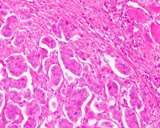
I have seen one case of what I called fibroadenomatous hyperplasia of the glands of the vestibule in a cat. The case was provided to me by Prof Selina Iussich (University of Turin) and Dr Barbara Banco (La Vallonea Veterinary Laboratory). The patient was an 11 year old cat with pyometra. There were multifocal small nodules in the vestibule, and histologically were papillary proliferations of both stroma and epithelium of the vestibular glands. A nodule of stroma was surrounded by epithelium, which itself was proliferative. In some places the epithelium alone was hyperplastic.

Vaginal polyp

A single vaginal polyp was recorded in the YB database

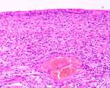
Figure : Vaginal polyp. The surface is ulcerated and there is edematous but normal tissue in the stroma.

Miscellaneous

Atresia

Nomura et al (1997) reports on a single case of vaginal atresia in a cat. There was a transverse septum present between the caudal and cranial portion of the vagina. The uterus and vagina proximal to the septum was distended with red brown fluid.

Nomura K, Koreeda T, Kawata M, Shiraishi Y. (1997) Vaginal atresia with transverse septum in a cat. J Vet Med Sci. 59(11): 1045-1048.

Laceration/Trauma (parturition injuries)

Bloom (1954) reports that tears during parturition are rare.

Bloom F (1954) Pathology of the dog and cat - The genitourinary system, with clinical considerations. American Veterinary Publications, Inc, Evanston Illinois. p320.


Vaginal Prolapse

McKelvey et al (2015) reported on a Maine Coon cat that developed a vaginal prolapse at 54 days of gestation. It was reduced and caesarian section was later performed to deliver the kittens.


McKelvey KA, Beachler TM, Ferris KK, Diaw M, Vasgaard JM, Bailey CS. Vaginal prolapse in a pregnant Maine coon cat: a case report. J Small Anim Pract. 2015l ;56: 473-475..


Rectovaginal fistula

Suess et al (1992) reports 3 kittens with atresia ani that developed rectovaginal fistulae.

Suess RP Jr, Martin RA, Moon ML, Dallman MJ. (1992) Rectovaginal fistula with atresia ani in three kittens. Cornell Vet. 82(2):141-153.


Vagino-ureteral fistula

Allen and Webbon (1980) report 2 cases of vagino ureteral fistulae. These were assumed to be acquired after ovariohysterectomy as the incontinence only began after surgery.


Allen WE, Webbon PM. (1980) Two cases of urinary incontinence in cats associated with acquired vagino-ureteral fistula. J Small Anim Pract. 21(7): 367-371.