This page is part of the site called Surgical Pathology of the Feline Female Reproductive Tract by

Dr Rob Foster
OVC Pathobiology
University of Guelph

Disease of the Feline Uterus

Table of Contents


General considerations

Lesions of the uterus of cats are common. The following table gives some indication of the occurrence of lesions and what to expect in submissions to a surgical pathology service.


Hyperplasia

Endometrial hyperplasia

65

Endometrial polyp

18

Adenomyosis

7
Endometritis/pyometra
40
Segmental aplasia
15
Remnants and duplications
11

Neoplasia

leiomyoma
7
endometrial stromal tumour
1

leiomyosarcoma

1
Mucometra/hydrometra
6
Serosal inclusion cysts
2
Rupture
1
Torsion
2
Haemosiderosis
1
 

Endometrial hyperplasia and associated diseases

There are several conditions that are inextricably linked to endometrial hyperplasia, and they include polyps and endometritis and pyometra. These conditions together make up the majority of the diseases with which a surgical pathologist will have to deal.

Cats have a propensity to develop a dramatic hyperplasia and changes that some think are atypia. Some even think this change is neoplastic and thus there are reports of diffuse adenocarcinoma of the uterus. I deal with these in the section on pseudopregnancy and endometrial hyperplasia.

Cystic endometrial hyperplasia

Cystic endometrial hyperplasia has been reviewed by several authors (Agudelo 2004). Cystic endometrial hyperplasia in the cat has almost always been thought to be similar to the disease in the bitch, but this may not be so. There are sufficient differences both in the disease and in the normal physiology of the 2 species to suggest they are not as similar as suspected.

Pathogenesis

Normal cyclic changes in the endometrium are under the control of ovarian steroidal hormones including oestrogen and progesterone. Physiological hyperplasia of the endometrium occurs during the luteal phase of the cycle and subsequent atrophy occurs during anoestrus. Ovarian androgens, produced by the theca interna may also play a role, and, while their function and control is not known, ovarian interstitial endocrine cells may play a role in this (Perez et al 1999). Oestrogen have a 'priming' function that allows the cells to respond fully to progesterone. Progesterone, though, alone will cause endometrial hyperplasia (Thornton and Kear 1967, Boomsma et al 1982, Chatdarong et al 2005)

Perez et al (1999) reported that feral queens did not have cystic endometrial hyperplasia, and their serum oestrogen concentration was lower during anoestrus than colony-reared queens. Feral queens also have more interstitial endocrine cells of the ovary. The control mechanisms involved are not known. The reproductive status of the feral cats, apart from them being mature animals, was unknown either.

Correlating concentrations of oestrogen and progesterone with degrees of hyperplasia, or with hyperplasia in general has been problematic, suggesting that local factors such as receptor concentration or the concentration of local endometrial growth factors may be more important. Boomsma et al (1997) examined feline endometrium for the presence of transforming growth factor, epidermal growth factor (EGF) and EGF receptor, and found them in the epithelium and stroma. Misirlioglu et al (2006) examined 10 normal cats and 20 cats with cystic endometrial hyperplasia and pyometra. They found that the endometrial hyperplasia group could be divided into 2 subgroups - those with mild hyperplasia and those with marked hyperplasia. They found that oestrogen receptor (ER) expression was lower in the mild hyperplasia group and higher in marked hyperplasia. c-erb-2 oncoprotein expression was also increased in both hyperplasia groups, as compared to controls. Progesterone receptors were lower in the hyperplastic groups than in controls.

Dow (1962) reported on 91 female cats with endometrial hyperplasia, uterine polyps and endometritis and pyometra. There were 20 cats with hyperplasia but no evidence of inflammation (9 also had polyps), 29 cats with endometrial hyperplasia and acute endometritis and pyometra (2 had polyps), 10 had 'subacute endometritis', and 22 had chronic endometritis While many affected cats had corpora lutea, some did not. The assumption is therefore that cats with endometrial hyperplasia are more prone to developing infection and inflammation.

Potter et al (1991) examined 79 queens with either clinical disease or lesions found at surgery or necropsy. 31 queens had clinically silent lesions of the uterus. 43 cats had never been bred, and 25 had clinical signs. All 79 cats had endometrial hyperplasia, 30 had pyometra, 10 had endometritis only, 21 had uterine polyps, and 5 had adenomyosis. 11 had cystic follicles, 6 had parovarian cysts, and 11 had cystic rete ovarii. Those with endometreal hyperplasia usually had some degree of uterine luminal dilation. They concluded that endometrial hyperplasia was seen mostly in older cats (>5yrs) and was a function of age in cats. Only 25 of the 79 cats with endometrial hyperplasia had a corpus luteum, so they surmised that hyperplasia was a function of oestrogen stimulation and not progesterone.

Based on these and subsequent findings, the general pathogenesis is assumed to be that the development of endometrial hyperplasia is related to hormonal and environmental factors. The effect of foreign material (i.e. infection) stimulating endometrial hyperplasia, as occurs in the dog, is unknown.

Macroscopic appearance

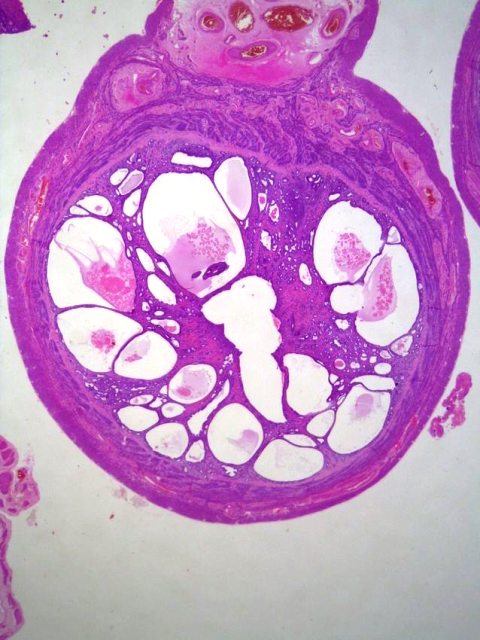
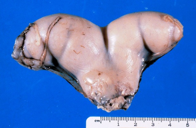
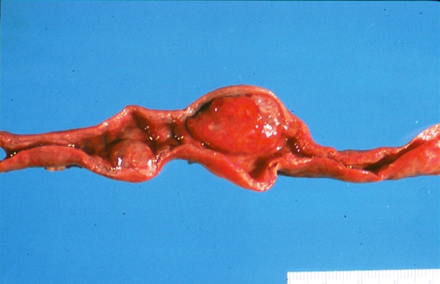
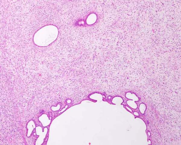
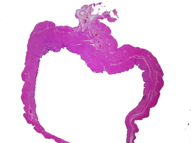
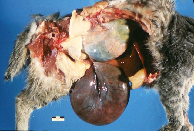
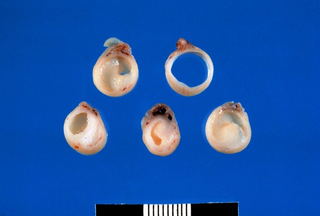
A uterus with endometrial hyperplasia is thicker and more convoluted than normal. In severe cases, this is especially visible on cross section where the corkscrew shape of the lumen is exaggerated and cysts are visible.

Figure : Endometrial hyperplasia. The normal corkscrew appearance of the endometrium is exaggerated and there is cystic dilation of the lumen (mucometra) and dramatic thickening of the endometrium.

Microscopic appearance

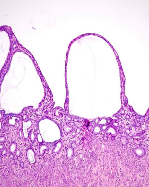
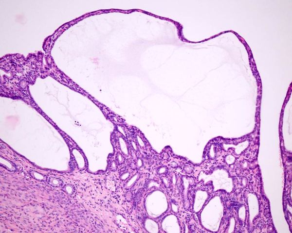
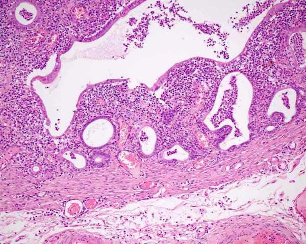
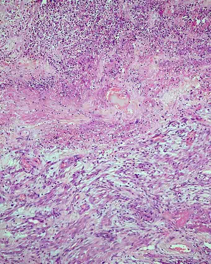
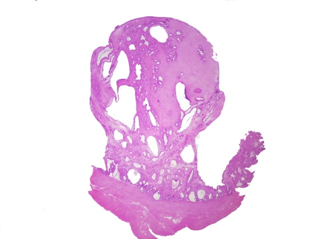
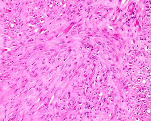
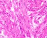
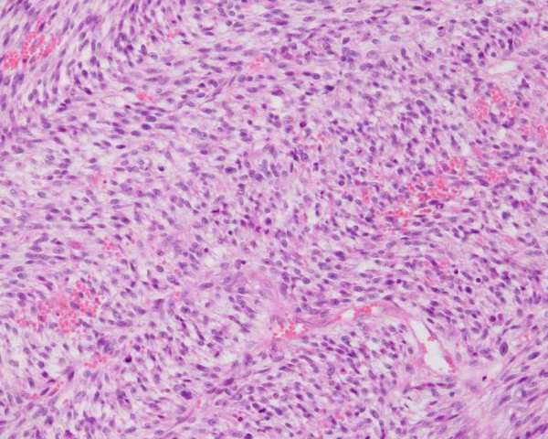
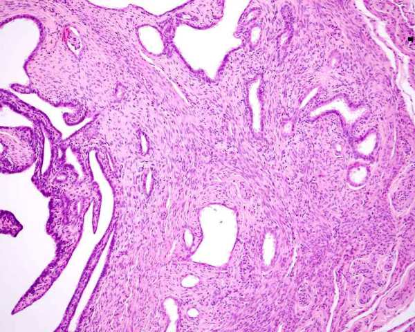
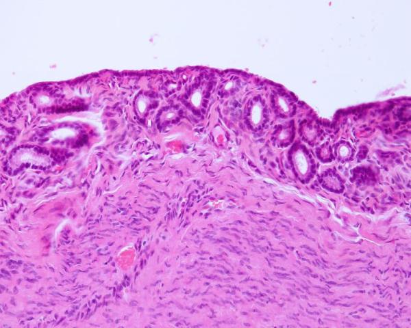
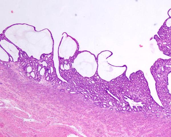
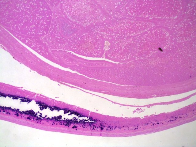
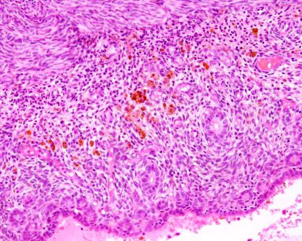
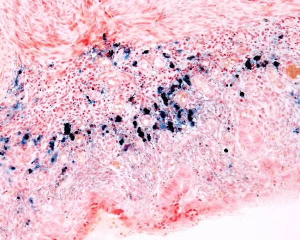
Histological changes in the endometrium with hyperplasia varies from mild to severe. Mild hyperplasia is a thickening of the endometrium with elongation of the glands. As this becomes more greater, the glands become disorganised and rather than straight, become convoluted so that the cross sections vary. The stroma becomes more prominent. There is dilation of the glands with the formation of variably sized cysts.

Figure : Cystic endometrial hyperplasia.

Agudelo CF. (2005) Cystic endometrial hyperplasia-pyometra complex in cats. A review. Vet Q. 27(4):173-182.

Boomsma RA, Jaffe RC, Verhage HG (1982) The uterine progestational response to cats: changes in morphology and progesterone receptors during chronic administration of progesterone to estradiol-primed and nonprimed animals. Biol Reprod 26: 511-521.

Boomsma RA, Mavrogianis PA, Verhage HG. (1997) Immunocytochemical localization of transforming growth factor a, epidermal growth factor and epidermal growth factor receptor in the cat endometrium and placenta. Histochemical J 29:495-504.

Chatdarong K, Rungsipipat A, Axner E, Linde Forsberg C. (2005) Hysterographic appearance and uterine histology at different stages of the reproductive cycle and after progestagen treatment in the domestic cat. Theriogenology. 64(1):12-29.

Dow C (1962). The cystic endometrial hyperplasia - pyometra complex in the cat. Vet Rec 74: 141-147.

Misirlioglu D, Nak D, Sevimli A, Nak Y, Ozyigit MO, Akkoc A, Cangul IT. (2006) Steroid receptor expression and HER-2/neu (c-erbB-2) oncoprotein in the uterus of cats with cystic endometrial hyperplasia-pyometra complex. J Vet Med A Physiol Pathol Clin Med. 53(5):225-229.

Perez JF, Conley AJ, Dieter JA, Sanz-Ortega J, Lasley BL (1999) Studies on the origin of ovarian interstitial tissue and the incidence of endometrial hyperplasia in domestic and feral cats. Gen Comp Endocrinol. 116(1):10-20.

Potter K, Hancock DH, Gallina AM (1991) Clinical and pathological features of endometrial hyperplasia, pyometra and endometritis in cats: 79 cases (1980-1985). J Am Vet Med Assoc. 198(8):1427-1431.

Thornton DAK, Kear M (1967). Uterine cystic hyperplasia in a siamese cat following treatment with medroxyprogesterone. Vet Rec 80: 380-381.

Endometritis and pyometra

Inflammation of the endometrium is endometritis. Pyometra is the filling of the uterus with pus.

Infection of the uterus may be more common than realised and not all infection leads to inflammation. Clemetson and Ward (1990) found bacteria in the uteri of 2 out of 29 cats sampled. Most cats have bacteria in the vagina, and ascending infection when the cervix is open at oestrus and especially at mating, is probably common. Natural protective mechanisms such as uterine contraction and fluid and mucus production eliminates infection in most circumstances. The uterus is prone to maintain infection if the nonspecific mechanisms fail, and especially during the dioestral phase of the cycle. The widely held belief that endometrial hyperplasia predisposes to infection and maintenance of infection may well be true, but there are no experiments to eliminate the alternate hypothesis that infection stimulates hyperplasia, as has been shown in dogs.

Clemetson LL, Ward ACS (1990) Bacterial flora of the vagina and uterus of healthy cats. J Amer Vet Med Assoc 196: 902-906.

Macroscopic changes

Endometrial hyperplasia is invariably present in cases of endometritis and pyometra, but the severity of the hyperplasia varies (Misirlioglu et al 2006). The macroscopic changes of endometritis are similar to those of endometrial hyperplasia, with the exception that there may be an exudate reassembling pus. When pyometra is present, the lumen of the uterus is distended and filled with pus. This can be up to 1.5 litres!

Figure : Pyometra.

Microscopic changes

The histological lesions begin with a migration of neutrophils into the glands and the lumen. Lymphocytes and plasma cells will form in the stroma of the endometrium. With a greater severity, particularly if the bacteria are toxigenic, there will be erosion and ulceration of the endometrium. Some cases just continue to accumulate neutrophils and the lumen will be tremendously dilated. The endometrium will not be so severely affected, and in some animals, squamous metaplasia will develop. The wall may become atrophic.


Figure : Suppurative endometritis. There is a background of cystic endometrial hyperplasia and neutrophils in the lumen and within dilated glands, and lymphocytes and plasma cells in the stroma.

Figure : Metritis. The endometrium is missing and ulcerated. The exudate (upper) merges with the myometrium (lower)

Figure : Pyometra. The lumen is dilated. The luminal epithelial cells are stratified squamous in type, the glands appear to be atrophic and the stroma of the endometrium is prominent.

Kenney KJ, Matthiesen DT, Brown NO, Bradley RL (1987) Pyometra in cats: 183 cases (1979-1984). J Am Vet Med Assoc. 191(9):1130-1132.

Endometrial polyps

Endometrial polyps are believed to develop from endometrial hyperplasia (Gelberg and McEntee 1984), and there is always some degree of endometrial hyperplasia.

In general there are 3 forms of endometrial polyps - those with both epithelium and stroma (endometrial polyp), stromal predominant (endometrial stromal polyp), and hypercellular stromal (hypercellular endometrial stromal polyp)

Gelberg and McEntee (1984) provides a detailed report on 14 cats with hyperplastic endometrial polyps. They suggested that the polyps developed from endometrial hyperplasia. Other authors have mentioned the finding of endometrial polyps. Dow (1962) reported on 91 female cats with endometrial hyperplasia, uterine polyps and endometritis and pyometra. There were 20 cats with hyperplasia and 9 had polyps, 29 cats had endometrial hyperplasia and acute endometritis and pyometra, and 2 had polyps, for a total of 11 cats with polyps. Potter et al (1991) examined 79 queens with either clinical disease or lesions found at surgery or necropsy. All cats had endometrial hyperplasia, 30 had pyometra, 10 had endometritis only, and 21 had uterine polyps.

I had access to 18 cases of endometrial polyps.

Macroscopic findings

Although polyps can be microscopic in size, most are recognised as swellings of the uterus. They may be single or multiple and occur anywhere in the uterus (Bloom 1954). They should have a pedicle, but some have a broad base. Most are solitary and, when the uterus is opened, appear as a nodule on a stalk. Intussusception or prolapse of the polyp can occur.

Figure : Uterine polyps. The uterus has been opened to show the 2 intraluminal polyps.

Microscopic findings

polyps are composed of endometrial tissue identical to surrounding endometrium. They usually are composed of an outer layer of epithelium and stroma within which are distended uterine glands. In all locations, the cells, be they epithelium or stroma, are well differentiated, and do not have dysplastic features. The microscopic findings are identical to those of cystic endometrial hyperplasia.

Figure : Endometrial polyp, subgross. The myometrium is visible at the bottom, and attached to it by a broad stalk is a polyp.

Dow C (1962). The cystic endometrial hyperplasia - pyometra complex in the cat. Vet Rec 74: 141-147.

Gelberg HB, McEntee K (1984) Hyperplastic endometrial polyps in the dog and cat. Vet Pathol 21: 570-573.

Potter K, Hancock DH, Gallina AM (1991) Clinical and pathological features of endometrial hyperplasia, pyometra and endometritis in cats: 79 cases (1980-1985). J Am Vet Med Assoc. 198(8):1427-1431. .

Pseudopregnancy and endometrial hyperplasia

Pseudopregnancy is divided into 'overt' and 'covert' conditions. Clinical pseudopregnancy is when the animal develops the physical (and behavioural) attributes of pregnancy without being pregnant. This can include lactation. Clinical manifestations of pseudopregnancy are not common in cats. Covert or subclinical pseudopregnancy is the period when a cat develops a corpus luteum (noncopulatory ovulation) or does not conceive after mating. In the literature, this is the most common use of the word pseudopregnancy. Foster and Hisaw (1935) studied this subclinical pseudopregnancy in cats by experimental induction. They examined the uterus after stimulating oestrus and ovulation with pituitary hormones. The endometrium became thicker, and there was a slight dilation of the glands and hyperplasia of the epithelium.

There is very little written about the macroscopic or microscopic features of animals with clinical pseudopregnancy.

Cats do develop pseudopregnancy when they ovulate but either dont mate or have a sterile mating. They can develop a distinct region of endometrial hyperplasia where there is marked hyperchromasia of the epithelium, bi or multinucleation and branching of the lumens or region between fronds. I suspect some people think this is endometrial carcinoma!

Suzuki et al (2021) reported on cats with what they called atypical endometrial hyperplasia. They diagnosed the lesions as noninvasive or early stage adenocarcinoma!!! Their photomicrographs are of a lesion of this type - the feline uterus, even in young cats, can develop hyperplasia that appears 'atypical'. Their cases had a good outcome. 3 had pyometra, and their ages were 11 months to 13 years.


Boomsma RA1, Mavrogianis PA, Verhage HG.Changes in endometrial and placental protein synthesis and morphology during pregnancy and pseudopregnancy in the cat. Biol Reprod. 1991; 44(2): 345-356.

Foster MA, Hisaw FL (1935) Experimental ovulation and the resulting pseudopregnancy in anoestrous cats. Anatomic Rec 62: 75-93

Suzuki S, Kitamura H, Hayashi K, Nakashima T, Okamura M, Shirai K, Kariya T, Endometrial disease in six cats with clinical and histopathological features resembling atypical endometrial hyperplasia in humans. J Comp Pathol 2021 189: 45-51. *

Uterine Neoplasia

Traditional classification schemes of neoplastic disease often focus on whether the pattern of differentiation is epithelial, mesenchymal, round cell or other. This type of classification works well in the uterus, however, distribution of the lesions is an all important factor, so this is the approach used here.

Myometrial neoplasia

Smooth muscle tumor (Leiomyoma)

The most common neoplasm of the wall of the uterus in the cat is reported to be the leiomyoma. The expected appearance and histological features of leiomyoma are identical to leiomyomas in other locations. They are usually well circumscribed lesions that blend with the adjacent smooth muscle. They are composed of tough white tissue that histologically is a mesenchymal tumour composed of cells arranged in streams, bundles and fasicles and which have well defined cytoplasmic boundaries, abundant eosinophilic cytoplasm and oval vesicular nuclei. No or only rare mitoses are seen. Some can have abundant fibrous tissue, or myxomatous tissue and may be designated fibroleiomyoma and fibromyxoleiomyoma and any variants!

Immunohistochemistry should show these to be smooth muscle actin positive, as well as vimentin positive, although in some smooth muscle tumours, vimentin can be negative. Miller et al (2003) report them to be estrogen receptor positive.

Bloom (1954) reports seeing feline uterine leiomyomas. Papparello and Roperto (1984) report seeing them. McEntee (1994) reports seeing uterine leiomyomas in 12 cats. There are 7 in the YB database.

Figure : Histology of a leiomyoma of uterus. Cells resemble smooth muscle, are relatively uniform and had no mitoses.

Bloom F (1954) Pathology of the dog and cat - The genitourinary system, with clinical considerations. American Veterinary Publications, Inc, Evanston Illinois. p 368-371

McEntee K (1990) Reproductive Pathology of Domestic Mammals. Academic Press. p184.

Miller et al (2003) report on 13 cats with uterine neoplasia. 3 were leiomyomas, and one was a leiomyosarcoma. No metastases were seen.

Papparella S, Roperto F. (1984) Spontaneous uterine tumors in three cats. Vet Pathol. 21(2): 257-258.

Smooth muscle tumor (Leiomyosarcoma)

The malignant counterpart of the uterine leiomyoma is the leiomyosarcoma. There are no published criteria that can be applied with any consistency and none that are correlated with outcome. As a result, many and perhaps all reported leiomyosarcomas are benign.

They are reported to have a similar gross appearance to leiomyomas, but may be larger, multinodular and metastatic. Histologically they have dysplastic features including marked anisokaryosis, numerous mitoses, and regions of necrosis. Reports are sparce for these with Cooper et al (2006) and Miller et al (2003) reporting one case each. The presence of metastatis, if present, will confirm their malignancy!

Immunohistochemistry should show them to be smooth muscle actin positive. The one case of Miller et al (2003) was Estrogen Receptor negative. The metastatic neoplasm reported by Cooper et al (2006) expressed caldesmon, smooth muscle actin, and estrogen receptor but was negative for desmin.

There is 1 in the YB database.

Figure : Histology of leiomyosarcoma of myometrium. There are anaplastic changes and numerous mitoses.

Cooper TK, Ronnett BM, Ruben DS, Zink MC. (2006) Uterine myxoid leiomyosarcoma with widespread metastases in a cat. Vet Pathol. 43(4):552-556.

Miller MA, Ramos-Vara JA, Dickerson MF, Johnson GC, Pace LW, Kreeger JM, Turnquist SE, Turk JR. (2003) Uterine neoplasia in 13 cats. J Vet Diagn Invest. 15(6):515-522.

Endometrial neoplasia

The normal endometrium has epithelial and stromal components, so neoplasia involving epithelial only, stromal only or combinations are seen. All are rare, but epithelial tumours appear to be the more common. The features of each are similar to those see in other species.

Endometrial adenocarcinoma

There are multiple reports, often single case reports, of endometrial adenocarcinoma in the cat. These reports are more numerous than other neoplasms of the endometrium and makes adenocarcinoma the most likely neoplasm to be encountered. Having said this, endometrial carcinoma is exceedingly rare and cases may be hyperplastic lesions (see pseudopregnancy and Endometrial hyperplasia above). Carcinoma is justified if there is distinct invasion of the myometrium or metastasis.

Single examples are reported by Preiser (1964), Belter et al (1968), Norris et al (1969), O'Rourke and Geib (1970), Papparella and Roperto (1984), Martin de las Mulas et al (1995), .

Meier (1956) reports on 2 cases, both of which were metastatic. Sontas et al (2012) reported on 2 cases, neither of which had subsequent clinical signs.

Miller et al (2003) report on 13 cats with uterine neoplasia. 8 were adenocarcinomas, 3 had peritoneal carcinomatosis, and one had pulmonary metastases.

Gil da Costa et al (2009) performed an immunohistochemical study on 8 endometrial carcinomas using many stains including pancytokeratins AE1 and AE3, cytokeratin-14, vimentin, a-actin, cyclo-oxygenase-2, E-cadherin, b-catenin, the progesterone receptor, the oestrogen receptor and caveolin-1. One of the cats had intravascular invasion. Metastasis was not recorded. Of the 18 reported with followup, 8 were metastatic.

Sontas et al (2013) reported two cases of endometrial carcinoma, both of which were suspected of having pyometra. Both were young cats, and based on a follow-up of about 21 and 23 months, no metastases occurred.

Payan-Carreira et al (2013) reported 4 cases in young queens. These were incidental findings but 3 of the 4 cats had concurrent pyometra. No metastases were identified, although the followups were short.

Saraiva et al (2015) examined 24 cases of endometrial adenocarcinomas using immunohistochemistry for ER-α, PR, Ki-67, CK7 and CK20. They found that ER and PR were lost as compared to normal endometria and CK7 and CK20 were retained. Ki67 increased. In their cases, myometrial invasion was observed in 16 of the 24, and vascular invasion was in 3 of 24. Serosal involvement was seen in 1 case. Saraiva et al (2016) examined expression of c-erbB-2 in normal endometrium and in adenocarcinomas. It is expressed in normal endometrium and in 20 of 34 cases of adenocarcinoma.

Suzuki et al (2021) diagnosed noninvasive or early stage adenocarcinoma in 6 cats with atypical lesion of the uterus. (see Pseudopregnany and endometrial hyperplasia above). None were clinically significant.


Belter LF, Crawford EM, Bates HR. (1968) Endometrial adenocarcinoma in a cat. Pathol Vet. 5(5): 429-431.

Gil da Costa RM, Santos M, Amorim I, Lopes C, Dias Pereira P, Faustino AM. (2009) An Immunohistochemical Study of Feline Endometrial Adenocarcinoma J Comp Path 140: 254-259

Martin de las Mulas J, Espinosa de los Monteros A, Carrasco L, van Niel M, Fernandez A. (1995) Immunohistochemical distribution pattern of intermediate filament proteins in 50 feline neoplasms. Vet Pathol. 32(6): 692-701.

Meier H (1956). Carcinoma of the uterus in the cat: two cases. Cornell vet 46: 188-200. as quoted by McEntee (1990)

Miller MA, Ramos-Vara JA, Dickerson MF, Johnson GC, Pace LW, Kreeger JM, Turnquist SE, Turk JR. (2003) Uterine neoplasia in 13 cats. J Vet Diagn Invest 15: 575-522.

O'Rourke MD, Geib LW. (1970) Endometrial adenocarcinoma in a cat. Cornell Vet 60(4): 598-604.

Papparella S, Roperto F. (1984) Spontaneous uterine tumors in three cats. Vet Pathol 21: 257-258.

Payan-Carreira R, Saraiva1 AL, Santos T, Vilhena H, Sousa A, Santos C, Pires MA (2013) Feline Endometrial Adenocarcinoma in females <1 Year Old: A Description of Four Cases. Reprod Dom Anim 2013; 48: c70-c77

Prieser H (1964) Endometrial carcinoma in a cat. Pathol Vet 1: 485-490.

Saraiva AL, Payan-Carreira R, Gärtner F, Fortuna da Cunha MR, Rêma A, Faria F, Lourenço LM, Pires Mdos A. An immunohistochemical study on the expression of sex steroid receptors, Ki-67 and cytokeratins 7 and 20 in feline endometrial adenocarcinomas. BMC Vet Res 2015; 11: 204.

Saraiva AL, Payan-Carreira R, Gärtner F, Faria F, Lourenço LM, Pires MA. Changes in c-erbB-2 immunoexpression in feline endometrial adenocarcinomas. Reprod Domest Anim 2016; 51: 33-39.

Sontas BH, Erdogan Ö, Apaydin Enginler SÖ, Turna Yilmaz Ö, Şennazli G, Ekici H (2013) Endometrial adenocarcinoma in two young queens. J Small Anim Pract 2013, 54: 156–159.

Suzuki S, Kitamura H, Hayashi K, Nakashima T, Okamura M, Shirai K, Kariya T, Endometrial disease in six cats with clinical and histopathological features resembling atypical endometrial hyperplasia in humans. J Comp Pathol 2021 189: 45-51. *


Endometrial stromal tumors

Neoplasms that phenotypically are stromal, are very rare in cats. They form nodules of regional thickenings of the endometrium and push aside, or focally invade the myometrium. Endometrial glands may be trapped within the neoplasm. They are stromal in type with spindle shaped cells in a myxomatous matrix that are arranged in a haphazard arrangement.

Three variants are recognised in humans and similar types occur in cats. They are endometrial stromal nodule - a well circumscribed expansile lesion, endometrial stromal sarcoma - low grade - an infiltrative stromal tumour, and endometrial sarcoma - high grade - which is anaplastic and infiltrative.

Papparella and Roperto (1984) report 3 cats with uterine tumors. 1 was a 'submucosal fibroma'.

Sorribas CE (1987) reports on a cat with a 'submucous fibroma'.

Sato et al (2007) reports on a cat with an endometrial stromal sarcoma. There was smooth muscle differentiation, and these and one half of the stromal cells stained for smooth muscle with caldesmon and desmin . They also report the presence of epithelium within the stroma and they also considered this to also be neoplastic. There were intravascular neoplastic cells and metastases in the lungs.

Hirota et al (2023) reported on one case. It was multifocal. There was local invasion, vascular invasion, and high grade. Some cells were CK positive, but they were SMA, Desmin and IBA1 negative. Vimentin was positive.

There is one endometrial stromal tumour in the YB database and it was an endometrial stromal sarcoma - low grade.

Figure : Endometrial stromal sarcoma. The neoplasm was infiltrative of the endometrium (upper photograph), did not appear to directly affect the myometrium, had areas of necrosis (middle photograph) and was cellular with numerous mitoses.

Hirota T, Yonemaru K, Hattori M, Murakami M, Sakai H, Hirata A. Highly malignant endometrial stromal sarcoma in a cat. J Comp Pathol. 2023; 208: 11-14.

Papparella S, Roperto F. (1984) Spontaneous uterine tumors in three cats. Vet Pathol 21: 257-258.

Sato T, Maeda H, Suzuki A, Shibuya H, Sakata A, Shirai W. (2007) Endometrial stromal sarcoma with smooth muscle and glandular differentiation of the feline uterus. Vet Pathol 44: 379-382.

Sorribas CE (1987) reports on a cat with a 'submucous fibroma'.

Mixed endometrial tumors (mesodermal tumour/Carcinosarcoma/Mixed Muellerian tumour/MMT)

Mixed neoplasms of the endometrium have both epithelial and stromal elements, and either can be histologically benign or malignant. They are named according to the elements present, with the epithelial component first.

Evans and Grant (1977) report one cat with a metastatic neoplasm involving the uterus and which regrew from the uterine stump. The mass had a carcinomatous and sarcomatous component.

Papparella and Roperto (1984) report 3 cats with uterine tumors. 1 was a carcinosarcoma. It was metastatic.

Nicotina et al (2002) reports on 1 cat with a carcinosarcoma. It was widely metastatic throughout the thorax and abdomen.

Miller et al (2003) report on 13 cats with uterine neoplasia. 1 was an adenosarcoma.


Evans JG, Grant DI (1977) A mixed mesodermal tumour in the uterus of a cat. J Comp Path 87: 635-638.

Miller MA, Ramos-Vara JA, Dickerson MF, Johnson GC, Pace LW, Kreeger JM, Turnquist SE, Turk JR. (2003) Uterine neoplasia in 13 cats. J Vet Diagn Invest 15: 575-522.

Nicotina PA, Zanghi A, Catone G. (2002) Uterine malignant mixed Mullerian tumor (metaplastic carcinoma) in the cat: clinicopathologic features and proliferation indices. Vet Pathol 39: 158-160.

Papparella S, Roperto F. (1984) Spontaneous uterine tumors in three cats. Vet Pathol 21: 257-258.

Other uterine neoplasia

Haemangioma

Fukui and Matsude (1983) found a 11 cm well circumscribed, pedunculated, hemorrhagic mass that histologically was typical of a haemangioma in the right uterine horn of a cat.

Fukui K, Matsuda H. (1983) Uterine haemangioma in a cat. Vet Rec 113(16): 375.

Lymphoma

Lymphoma of the uterus is rarely reported.

Azakami et al (2016) reported a case of a high grade B cell lymphoma that presented as a mass in the uterine horn. The cat was treated with surgery and chemotherapy and died of complications 310 days later.

Conversy et al (2017) reported a case where there was a nodule in the uterus of an ovarectemised cat that was diagnosed as a T cell lymphoma. The cat developed a retrobulbar B cell lymphoma 19 months later.

Azakami D, Onozawa E, Miyabe M, Ochiai K, Michishita M, Hirano T, Momota Y, Ishioka K, Sako T. Primary T-cell high-grade lymphoma of the feline uterus. J Vet Med Sci. 2016 Jun 1;78(5):913-6

Conversy B, Freulon AL, Graille M. Focal uterine T-cell lymphoma in an ovariectomized cat. J Am Vet Med Assoc. 2017; 251: 1059-1063.


Adenomyosis

Adenomyosis of the uterus is the presence of endometrial tissue within the myometrium. The pathogenesis is unknown, and there is speculation as to how the endometrium comes to be within the myometrium. According to Bloom (1954), serial section of affected regions of the uterus show a connection to the endometrium. Endometrial hyperplasia is usually present. Some assume the endometrium 'grows' into the myometrium, but the stimulus or cause for such an invasion is not clear. Likewise, being forced into the myometrium under pressure is equally difficult to imagine. Having said this, McEntee (1990) reports seeing an artefact of fixation where endometrium appears in the myometrium in uteri fixed without being opened.

The disease is clinically silent and any signs are referable to endometrial hyperplasia.

Dow (1962) reports seeing on case in the uterine body. Potter et al (1991) examined 79 queens with either clinical disease or lesions found at surgery or necropsy. 5 had adenomyosis. Bulman-Flemming (2008) reported one case. There are 7 cases in the YB database.

Macroscopic findings

The gross lesions of adenomyosis are difficult to recognise grossly. It appears as though adenomyosis occurs in the uterine body, and if severe can cause a thickening of that region. The cut section will have small cystic structures in the myometrium.

Microscopic findings

Some cases of adenomyosis are identified only by the very observant, and even then, must be differentiated from sectioning artefact. Where the amount of endometrial tissue in the myometrium is extensive, the diagnosis is made easier. The endometrial tissue includes epithelium and stroma, and there is often hyperplasia of the elements, just like the adjacent endometrium. Cystic dilation of the epithelial components is common.

In the case documented by Bulman-Fleming (2008), there was supposed to be diffuse adenomyosis of all uterine horns and the body. This is most unusual. This could not be verified with the photograph published, as the sample appeared to have been cut longitudinally and parasagitally so that the appearance of glands in the muscular wall could have been artifactual as the endometrium of cats is a corkscrew shape and the sample may have been multiple views of the hypoplastic endometrium. We must rely on the author for the correct interpretation, however.

Figure : Uterine adenomyosis. The body of the uterus has endometrium within the myometrium (right). The endometrium is hyperplastic.

Figure : Adenomyosis. Uterine glands and stroma are within the myometrium.


Dow C (1962). The cystic endometrial hyperplasia - pyometra complex in the cat. Vet Rec 74: 141-147.

Bulman-Fleming J. (2008) A rare case of uterine adenomyosis in a Siamese cat. Can Vet J. 2008 49(7): 709–712.

Potter K, Hancock DH, Gallina AM (1991) Clinical and pathological features of endometrial hyperplasia, pyometra and endometritis in cats: 79 cases (1980-1985). J Am Vet Med Assoc. 198(8): 1427-1431.

Bloom F (1954) Pathology of the dog and cat - The genitourinary system, with clinical considerations. American Veterinary Publications, Inc, Evanston Illinois. p352

McEntee K (1990) Reproductive Pathology of Domestic Mammals. Academic Press. p171.

Mucometra/hydrometra

The accumulation of watery fluid within the uterus is hydrometra, and if the fluid is mucinous, it becomes mucometra. The contents in these conditions are acellular or poorly cellular.

In most conditions, the accumulation of fluid is the result of an obstruction to outflow. Cystic endometrial hyperplasia can contribute to this. Obstruction to outflow is seen in improper attempts to remove the uterus during ovariohysterectomy and leaving a portion of uterus behind (see ovarian remnant syndrome). The uterine remnant becomes cystically distended.

Obstruction at the cervix will cause the uterine body and both horns to distend. Endometrial polyps can cause a local obstruction and the uterus will distend proximal to the site. Segmental aplasia and uterine neoplasia are other potential causes. Those cases where obstruction is the cause would be expected to have atrophy of the endometrium due to pressure.

Hydrometra and mucometra are also seen in some cases of cystic endometrial hyperplasia, and varying degrees of dilation may be seen. In mild cases, the dilation is best recognised after transverse sectioning of the uterus (see cystic endometrial hyperplasia above). Marked dilation will require some obstructive situation.


Figure : Hydrometra. The uterus is distended and the lumen dilated. The mesometrium is uppermost.

Figure : Hydrometra with atrophy of the endometrium. A downstream obstruction would explain this change.

Figure : Mucometra with cystic endometrial hyperplasia

Figure : Endometrial hyperplasia in a cat with mucometra.


Cysts and embryonic remnants

The literature on cysts and embryonic remnants is sparse in the cat. Despite this, many submissions have remnants of embryonic ducts - there are 11 in the YB database.

The section on remnants of the mesonephric and paramesonephric systems for the ovary and uterine tube apply here as well. In the area of the uterus, there are several choices for cysts. They are:-


Serosal inclusion cysts

Serosal inclusion cysts are common in dogs, but they are very rare in cats. They are this walled fluid filled cystic structures on the serosal surface of the uterus. They are often multilocular and are attached by a thin stalk. They have a lining of thin squamous (flattened) cells that in some parts of the cyst may become cuboidal. The wall is this and compressed collagen. They are believed to arise when mesothelial cells become trapped in the serosa and continue to secrete, become hyperplastic and form the characteristic structures.

Godfrey and Silkstone (1998) found multiple cysts on the serosal surface of a multiparous queen.

Maya et al (2022) reported a case.

Ive seen at least 2 cases

Figure : Serosal inclusion cysts. This thin walled multicystic structure was attached to the serosa of the uterus.


Godfrey DR, Silkstone MA. (1998) Uterine serosal inclusion cysts in a cat. Vet Rec.142(24): 673.

Kaya MT, Kuruca N, Kanat M, Akca FB, Gulbahar MY. Uterine Serosal Inclusion Cysts with Greater Omentum Involvement in a Cat. J Comp Pathol. 2022; 199: 37-42.

Uterine Prolapse

Prolapse of the uterus is uncommon in the cat. Most cases are readily recognised clinically and the samples are not sent to a pathologist for examination.

Prolapse occurs after parturition, or in rare cases where an endometrial polyp has prolapsed and taken the uterus with it.

There are many individual case reports of uterine prolapse in cats. One unique one is Bigliardi et al (2014) where the uterus prolapsed through a perforation in the vagina after parturition.


Bigliardi E, Di Ianni F, Parmigiani E, Cantoni AM, Bresciani C. (2014) Complete uterine prolapse without uterine mucosal eversion in a queen. J Sm Anim Pract 2014;

Bruinsma DL. (1981) Feline uterine prolapse (a case report). Vet Med Small Anim Clin. 76(1):60.

Davies JE. (1978) Prolapsed uterus in the cat. Vet Rec 103(25):567.

Herbert CR. (1979) Prolapsed uterus in the cat. Vet Rec 104(2):42.

Maxson FB, Krausnick KE. (1969) Dystocia with uterine prolapse in a Siamese cat. Vet Med Small Anim Clin. 64(12):1065-1066.

van der Kolk FR.(1984) Uterine inversion and prolapse in a cat Tijdschr Diergeneeskd. 109(18):702-707.

Vanderhust SR. (1975) Bicornuate uterine prolapse in a cat/ a photographic essay. Vet Med Small Anim Clin. 70(6):681.

Vaughan L, McGluckin S (1993) Uterine prolapse in a cat. Vet Rec : 568

Wallace LJ, Henry JD Jr, Clifford JH. 1970 Manual reduction of uterine prolapse in a domestic cat. Vet Med Small Anim Clin. 65(6):595-596.

Uterine rupture / Ectopic pregnancy

Uterine rupture occurs only in pregnant animals, and often in advanced pregnancy. A traumatic incident is usually involved, although ischaema of the uterus with prolonged parturition in an oversized foetus is also possible.

A surgical pathologist may be sent a portion of the rupture site to ascertain if there is an underlying problem (like infarction) of the wall of the uterus. The other time a pathologist is involved is if a mass is removed from the abdomen that resembles a foetus.

Ectopic pregnancy is the condition in primates, humans in particular, where the foetus develops outside the uterus. Development is in the uterine tube, and eventually the foetus and membranes cause rupture of the uterine tube and pain and haemorrhage occurs. This is not reported in domestic mammals. What is reported is 'ectopic pregnany' where there has been previous rupture of the uterus an the foetus becomes ectopic. The foetus and membranes become mummified and are found as incidental masses in the abdomen.

There are individual case reports of rupture of the uterus and there is 1 case in the YB database. Individual reports of 'ectopic pregnancy' are reported. I have seen 2 cases of 'ectopic pregnancy'. One cat had been neutered 8 years previously and had a mummified term foetus removed from its abdomen when an abdominal mass was palpated.

Figure : Histology of "Ectopic Pregnancy". The wall of the foetus with membranes has a thick outer fibrous capsule and an inner material that is the necrotic remains of fatal membranes. The zonary placenta is visible in the upper photograph. The wall is often partially mineralised.


Bodle TJ. (1979) Ectopic pregnancy in a cat. N Z Vet J. 27(12): 279

Crownover RW, Yeargan GS Jr. (1976) Extra-uterine pregnancy in a spayed cat. Vet Med Small Anim Clin. 71(12): 1698-1699.

De Nooy PP. (1979) Extrauterine pregnancy and severe ascites in a cat. Vet Med Small Anim Clin. 74(3): 349-350.

Forbes NA. (1989) Ectopic pregnancy in a cat. Vet Rec. 124(22):595.

Hannon CA. (1981) Mummified fetuses in a cat. Mod Vet Pract. 62(2): 133-134.


Hansen JS. (1974) Ectopic pregnancy in a queen with one uterine horn and a urachal remnant.
Vet Med Small Anim Clin 69(9): 1135-1137

Hosgood G. (1989) Uterine rupture with intraperitoneal foetuses in a cat. Aust Vet Pract 19: 150-151.

King GJ, Amoroso EC (1983). Unusual phenomena during pregnancy in the cat and cow. Can J Comp Med 47: 379-381.

Knott CD (1989) Ectopic pregnancy in a cat. Vet Rec : 124(25):666


Johnston SD, Harish G, Stevens JB, Scheffler HG. (1983) Ectopic pregnancy with uterine horn encapsulation in a cat. J Am Vet Med Assoc.183(9): 1001-1002, 965.

Laube T. (1986) Primary and secondary extrauterine pregnancy in a cat. Tierarztl Prax. 1986;14(4):509-513.

Lofstedt R. (1989) Questions extrauterine development of fetuses. J Am Vet Med Assoc. 194(3):326-327.

Lucas X, Agut A, Ramis G, Belda E, Soler M. (2003) Uterine rupture in a cat. Vet Rec. 152(10): 301-302.

McKeating FJ. (1979) Ectopic pregnancy in a cat. 17: 104(11): 240-241.

Nack RA (2000). Theriogenology question of the Month. J Amer Vet Med Assoc 217: 182-184

Palmer NE (1989) Ectopic pregnancy in a cat. Vet Rec 125(1): 24

Thiel W, Knollner P, Weingartner E.(1988) A chance finding during the sterilization of a cat: a mummified fetus in the abdominal cavity. Tierarztl Prax. ;16(2):193-195.

Uterine torsion

Uterine torsion is a rare complication of pregnancy in cats. It involves one horn and the twist is at the junction between the body and one horn, and it involves the proximal end of the uterus or even the ovary. Affected cats may be found dead.

The affected portion is dark red black in colour from venous infarction.

Figure : Uterine torsion in a cat. The affected part of the uterus is red/black swollen (lower) as compared to the normal pregnant uterus (upper central).


Biller DS, Haibel GK. (1987) Torsion of the uterus in a cat. J Am Vet Med Assoc. 191(9): 1128-1129.

Manda JA (1986) Identifying uterine torsion in pregnant cats. Vet Med. p936-938.

Thilagar S, Yew YC, Dhaliwal GK, Toh I, Tong LL. Uterine horn torsion in a pregnant cat. Vet Rec. 157(18): 558-560.

v d Eijk PA, de Jong MF. (1981) Uterine torsion in a cat Tijdschr Diergeneeskd. 106(8): 410-411.

Young JD, Hillis GP, McKibbin ML. (1992) Uterine torsion in a cat. Feline Pract 20: 27-28.


Miscellaneous diseases of the uterus

Endometrial haemosiderosis

Haemosiderin laden macrophages are occasionally seen in the endometrium of postpartum animals, but widespread discolouration of the endometrium is unusual. One such case had no other indications of uterine disease, but the endometrium was a brown black colour and the endometrium contained a large amount of haemosiderin pigment. The cause is not known, but excessive uterine bleeding was likely.

Figure : Endometrial haemosiderosis. Large amounts of iron pigment is present in the stroma of the endometrium. Lower photograph is a Perl Prussian blue stain.