This page is part of the site called Surgical Pathology of the Feline Female Reproductive Tract by

Dr Rob Foster
OVC Pathobiology
University of Guelph

Disease of the Feline Ovary

Table of Contents


General considerations

Most pet female cats are spayed, and as a result, dont have the opportunity to get disease of the reproductive tract. The following table gives an indication of the types of submissions with which a surgical pathologist must deal (from Yager-Best Veterinary Surgical Pathology). Clearly, reoccurrence of estrus after neutering surgery is the most frequent reason for submission. Virtually all the other submissions were because something unusual was seen when the queen was spayed. Of all of the submissions, only one was supposed to be potentially malignant (a granulosa cell tumour).

Total submissions
141
Search for ovarian remnants
56
Remnants found
43
No remnants found
13
Normal ovaries submitted
19

Cystic structures

31

cystic rete ovarii

29
follicular cysts
2
inclusion cyst
1
Neoplasia
9
Sex cord stromal
5
Dysgerminoma
3
Teratoma
1
Hyperplasia of interstitial cells
1
Oophoritis
5
Ectopic adrenal
5
Hypoplasia
4
Distended lymphatics
2
Varix
1


Ovarian remnant syndrome

Examination for ovarian remnants is very common. Ovarian remnant syndrome is described in the general section on reproductive disease as a complication of ovariohysterectomy.

Ovarian and periovarian cysts

It is very difficult to positively identify the origin of cystic structures of or around the ovaries based on histology alone. Location of the cyst is extremely important. Dividing cysts into those in the ovary (ovarian cysts) and those outside the ovary (periovarian cysts) assists in determining this.

Ovarian cysts (within the ovary)

Cystic rete ovarii

These are very common in cats. and are detailed in Disorders of Sexual Development in cats

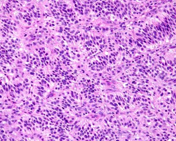
There are 3 parts to the rete ovarii - the intraovrian rete, the extraovarian rete, and a connecting rete. The rete are supposed to have a secretory activity. The intraovarian rete is supposed to be noncilited, whereas the connecting rete is lined by ciliated epithelial cells.

Cystic rete ovarii are when the rete ovarii become distened with fluid. It would be expected that the rete would enlarge with age, as the remnants are supposed to be secretory, and as an extension to this, all old cats should have cystic rete. This is not the case! They occur in young as well as old cats (McEntee 1990)

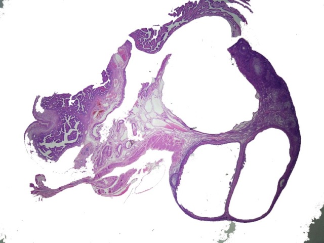
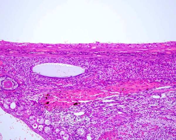
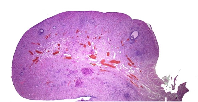
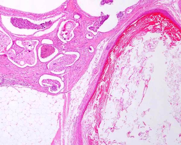
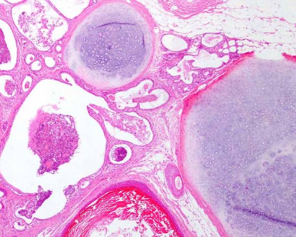
Rete cysts are thin walled structures that occur within the ovary and displace ovarian tissue peripherally. They can be up to 2.5 cm diameter. They are lined by a flattened single layer of epithelium. A thin layer of connective tissue is at the periphery, and this included compressed ovarian stroma.

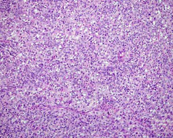
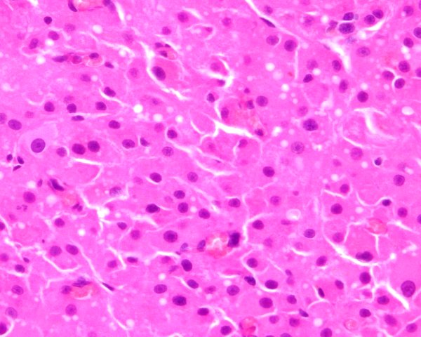
Gelbert et al (1984) described cystic rete ovarii in 20 cats. The cysts were within the ovary or at the tubal extremity of the ovary. The cyst lining was variable - they were single or multilayered, cuboidal to columnar, ciliated and nonciliated, or combinations of these.

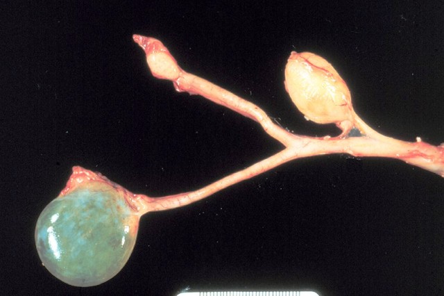
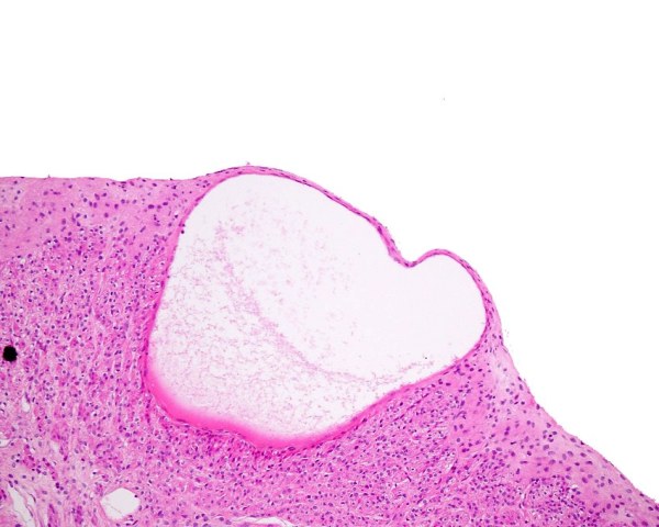
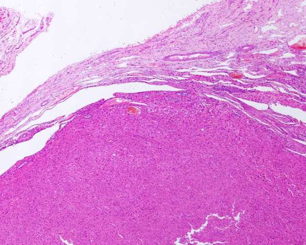
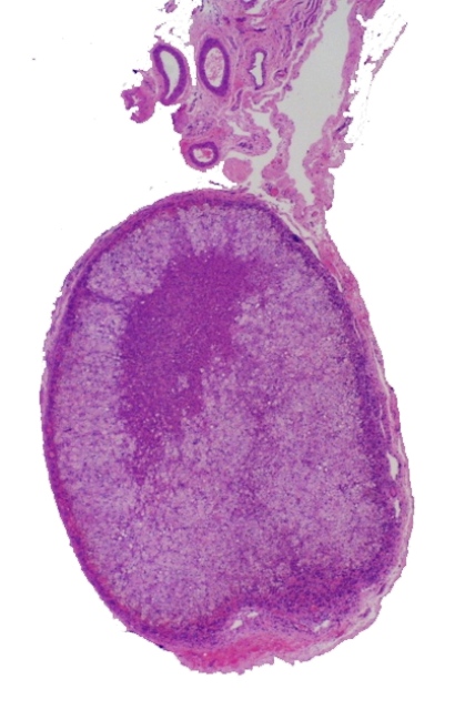
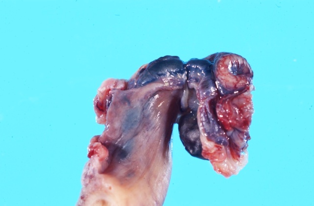
Figure : Unilateral cystic rete ovarii.

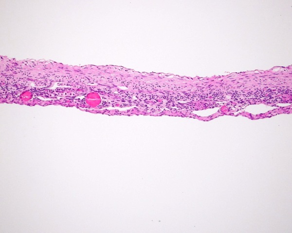
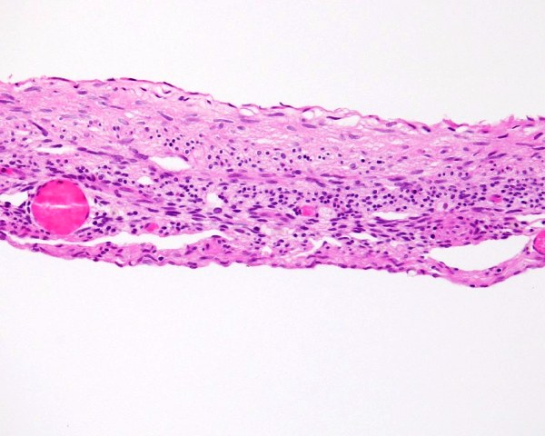
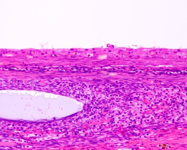
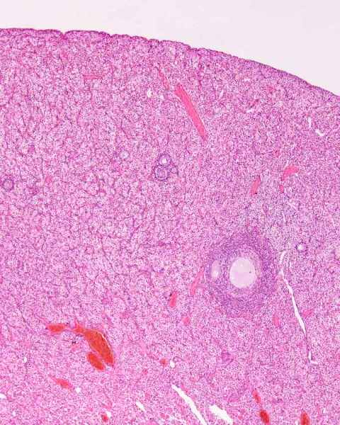
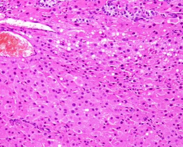
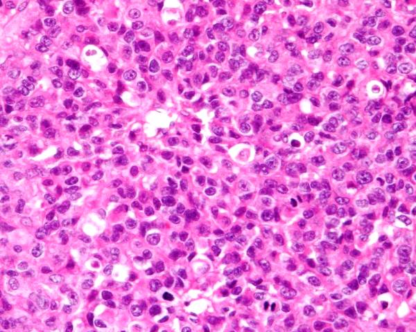
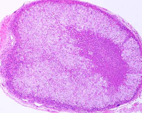
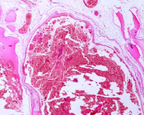
Figure : Cystic rete ovarii. Wall of cyst.

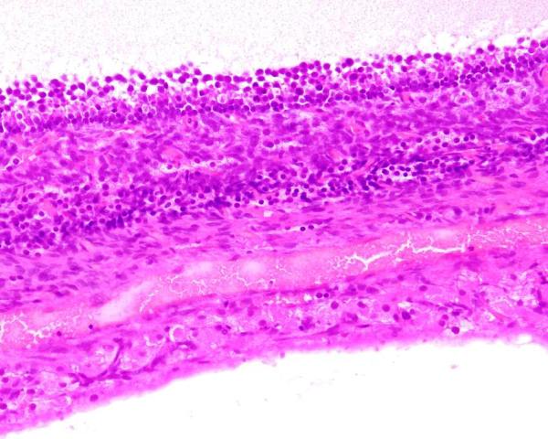
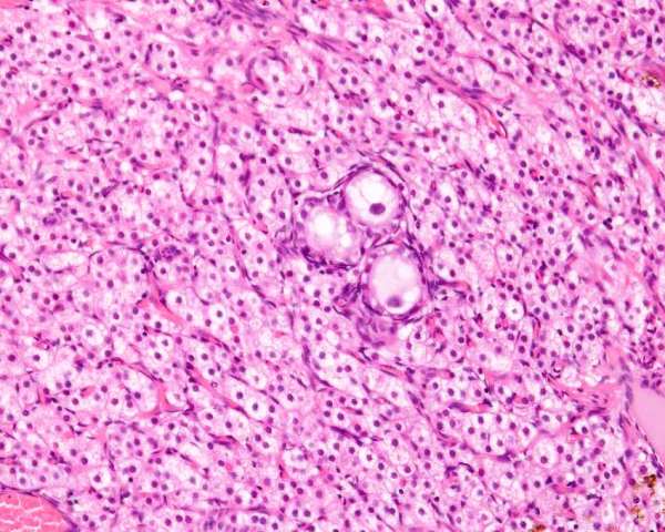
Figure : Cystic rete ovarii. Wall of cyst has this layer of epithelium then compressed collagen and ovarian stroma.

Gelberg HB, McEntee K, Heath EH (1984) Feline cystic rete ovarii. Vet Pathol 21: 304-307

McEntee K (1990) Reproductive Pathology of Domestic Mammals. Academic press p61

Cystic follicles

The normal follicle of the queen is supposed to be up to 2 to 3 mm diameter. Larger follicles are said to be cystic.

Cystic follicles have the same structure as normal follicles, with a layer of granulosa and thecal cells at the periphery. In other species they represent a failure of the follicles to ovulate. Little (nothing!) is known of them in cats.

The cystic follicles can be up to 7 mm diameter, and are frequently seen in ovarian remnants, presumably because they develop in an altered hormonal environment of increased pituitary gonadotrophins. Affected cats will have prolonged estrus or persistent estrus.

Figure : Subgross photograph of follicular cysts.

Figure : Ovary with follicular cyst (upper), stroma containing interstitial endocrine cells and ova (lower left)

Figure : Ovary with follicular cyst (upper), stroma containing interstitial endocrine cells and a secondary follicle.

Figure : Ovary with follicular cyst (upper) and stroma containing interstitial endocrine cells.

McEntee K (1990) Reproductive Pathology of Domestic Mammals. Academic press p57

Serosal inclusion cysts

An inclusion cyst is a cystic structure forms when there is trapping of mesothelium from the serosa in a region beneath the serosal surface. The cat does not have subsurface epithelial structures but cysts involving the surface of the ovary arise. They may form when a follicle ovulates and mesothelium enters the cavity. For most, there is no evidence of a cause. They appear as thin walled fluid filled cysts that are visible on the surface of the ovary, and project above the surface. Histologically, they are lined by a single thin layer of flattened cells that appear too be either squamous or low cuboidal.

.

Figure : Serosal inclusion cyst

Cystic corpus luteum

An otherwise normal corpus luteum with a large central cavity is a cystic corpus luteum. This is believed to occur when there is incomplete formation, perhaps from premature closure of the site of rupture of the follicle. McEntee (1990) has a photograph of a cystic corpus luteum in a cat.

McEntee K (1990) Reproductive Pathology of Domestic Mammals. Academic Press p59.

'Periovarian' cysts (outside the ovary)

Cysts that occur outside the ovary are commonly called (and according to McEntee 1990 inaccurately) parovarian cysts. Parovarian cysts are actually cysts of the epoophoron (parovarium) or cranial mesonephric tubules, this do not encompase all the cysts that occur outside the ovary. Cysts outside the ovary (periovarian!!) cysts arise from either remnants of the mesoneprhic (Wolffian) tubules or duct, or the paramesonephric (Mullerian) duct. While there are general guidelines about how to differentiate these, it is not easy. Location tends to be very important. McEntee (1990) used location as follows. Those between the fimbria and the ovary on the cranial extremity are remnants of the cranial mesonephric tubules. Those on the uterine or caudal pole of the ovary are remnants of the caudal mesonephric tubules. Ducts or cysts near the uterine tube are mesonephric duct remnants, and those on the serosal side of the fimbria of the uterine tube are remnants of the paramesonephric duct.

Histologically, paramesonephric duct remnants have a pseudostratified epithelium that may be partly ciliated. The nuclei of these remnants are about 50% larger than those of mesonephric origin. The mucosa forms papillae and is folded. No basement membrane is supposed to be present. There is a thin lining of smooth muscle or collagen. Remnants of mesonephric ducts have a simple columnar to cuboidal epithelium. Ciliated cells may be present.

Mesonephric remnants are very common in cats (Shehata 1974), but cysts are rare, and will be examined in more detail in the section on the uterine tube.

Shehata R. (1974) Mesonephric remnants in the female genital system of the domestic cat. Felis domestica. Acta Anat (Basel). 87(2):301-10.

Inflammation (oophoritis, ovaritis)

Inflammation of the ovary is called oophoritis (from ooophoron or ovary) or ovaritis. Oophoritis involving the stroma of the ovary is very rare, but perioophoritis secondary to ascending infection from salpyngitis is more common. It will be detailed further in diseases of the uterine tube

Ovarian enlargement

Interstitial endocrine cell hyperplasia

Ovarian hyperplasia is extremely rare and not reported. I have seen two cases of bilateral interstitial endocrine cell hyperplasia. Terminology is difficult here, as there will always be a dispute between what is hyperplastic and what is neoplastic. In this case, it is assumed the ovary appeared to be larger than normal (or the contralateral) ovary. Histologically there was expansion of the ovary with interstitial endocrine cells. Normal elements were separated rather than pushed to one pole or aside. The cells were uniform and typical of interstitial endocrine cells. While this case was called hyperplasia, a similar lesion was seen in another species that also had multiple metastases of the same cells! This case was called hyperplasia because of the diffuse nature of the infiltrates, and should be compared with interstitial cell neoplasia below.

Figure : Interstitial endocrine cell hyperplasia, with follicles isolated by diffuse expansion of interstitial endocrine cells

Ovarian neoplasia

General

Neoplasia of the ovary of cats is not common seen in surgical pathology practice. The types of ovarian neoplasms are categorises in a similar way to all gonadal neoplasms - being those of the gonadal stroma, germ cells or epithelium. While there are many potential subtypes, the usual ones and their occurrence are indicated in the table below.

 
Norris (1969)
Gelberg and McEntee (1985)
YB
Gonadal stromal tumours
6
13
4
Granulosa cell tumour
5
9
2
Interstitial cell tumour
1
5
2
Testicular sustentacular (Sertoli) cell tumour
   
1
Germ cell tumours      

dysgerminoma

1
6
3

teratoma

1
1
1
Epithelial tumours      
adenoma
 
1
 

carcinoma

1


Gelberg HB and McEntee K (1985) Feline ovarian neoplasms Vet Pathol 22: 572-576

Norris HJ et al 1969 Pathology of feline ovarian neoplasms. J Comp Path 97: 138-142

Sex cord - stromal (gonadostromal) tumors

Neoplasms of the ovary that phenotypically resemble the interstitial fibrous tissue, cells of the Graafian follicle or the interstitial endocrine cells are grouped into this category. It is not known for sure if all or some of these cells arise from the actual stroma of the gonad, from the mesonephric tubules, or from the embryonic coelomic lining (or combinations thereof)..

In general, these are the most common neoplasms of the feline ovary. They have the potential to produce steroidal hormones. Nothing is known of the pathogenesis (oncogenesis) of these neoplasms. They have malignant potential, but because they occur so infrequently, little is known of prognosis.

The macroscopic (surgical) appearance of sex cord - stromal neoplasms is dependent on the exact type, but they have the general 'stromal' appearance of being white to tan in colour, firm and multinodular. Some are multicystic.

Histologically their phenotype varies, and this variation can be within a neoplasm - some are uniformly of one type, but most have features of several subtypes. Most people either name them based on their predominant type, some call them mixed gonadal stromal tumours and others list the different types. The World Health Organisation, International Histological Classification of Tumors of Domestic Animals provides a further subclassification of these.

I suggest that the name of the neoplasm be sex cord - stromal tumour of ovary and the predominent type or variant be provided.

Granulosa cell tumours (sex cord - stromal tumour of ovary, granulosa cell tumour)

Granulosa cell tumours are the most common (see table above). These tumours resemble granulosa cells and of those reported about one third are metastatic (Gelberg and McEntee 1985). Granulosa cells are supposed to be round to oval cells with a clear or pale eosinophilic cytoplasm. The nuclei are hyperchromatic and round, and have distinct nucleoli. Theca cells are oval to spindle shaped with a vaculoated cytoplasm, or resemble fibroblasts. Most tumours have both phenotypes - the name given usually reflects the predominent type.

The arrangement of the cells varies also. Some form small (microfollicular pattern) or large (macrofollicular pattern) follicular patterns, or a diffuse pattern of cells. Those resembling testicular sustentacular (Sertoli) cell tumours of the testis are called testicular sustentacular cell tumours (sex cord - stromal tumour of ovary, testicular sustentacular (Sertoli) cell type).

A feature of granulosa cells is the formation of a rosette like structure with a central pale or eosinophilic fluid that are known as Call-Exner bodies. They are PAS postive. In feline tumours, Norris reports seeing Call-Exner bodies in 2 of 5 cats with granulosa cell tumours.

Immunohistochemical staining to confirm a tumour as a granulosa cell tumor is usually non required. Reports of staining in feline granulosa cell tumours is lacking. They should be cytokeratin negative, vimentin positive, inhibin positive, and may also stain with NSE and S100.

Norris et al (1969) reports on ovarian neoplasms in 10 cats. 5 cats had granulosa cell tumors. Call-Exner bodies were seen in 2. No mention of metastasis was made.

Figure : Sex cord - stromal tumour of ovary, granulosa cell tumour, diffuse pattern.


Figure : Sex cord - stromal tumour of ovary, granulosa cell tumour, diffuse pattern.

Figure : Sex cord - stromal tumour of ovary, testicular sustentacular cell pattern.

Aliakbrai S, Ivoghli B. (1979) Granulosa cell tumor in a cat. J Am Vet Med Assoc. 174(12):1306-1308.

Gelberg HB and McEntee K (1985) Feline ovarian neoplasms Vet Pathol 22: 572-576

Norris HJ et al 1969 Pathology of feline ovarian neoplasms. J Comp Path 97: 138-142


Thecoma (sex cord - stromal tumour of ovary, thecoma)

Theca cells are oval to spindle shaped with a vaculoated cytoplasm, or resemble fibroblasts. Neoplasms of this type resemble fibromas, but there is vacuolation of the cytoplasm with lipid. These are rare and not specifically reported in cats. Most would be grouped in with granulosa cell tumour.

Interstitial cell tumours (sex cord - stromal tumour of ovary, interstitial cell tumour)

Cats develop a proliferative condition wherein the interstitial cells of the ovary become greatly expanded in a fashion similar to gonadal hypertrophy of the equine foetus. This is described above under ovarian hyperplasia

Neoplasms of the ovary that are composed of round to polyhedral cells that have a vacuolated cytoplasm being either a single large vacuole or multiple fine granuoles, and a single central round nucleus or that resemble interstitial endocrine cells, adrenal cortex, interstitial cell tumours of the testis, or luteal cells are classified as interstitial cell tumours (Kennedy et al 1998).

Gelberg and McEntee (1985) report on 22 cats with ovarian neoplasms. 14 had gonadal stromal tumors of which 5 were interstitial cell tumors.

Norris et al (1969) reports on ovarian neoplasms in 10 cats. 1 cat had what they called a 'lipid-cell tumour', presumably a neoplasm of the interstitial endocrine cells. No mention of metastasis was made. This cat had virilization.

There are two in the YB database.

Figure : Histology of a feline ovarian interstitial cell tumour. This one resembles a luteoma

Gelberg HB and McEntee K (1985) Feline ovarian neoplasms Vet Pathol 22: 572-576

Kennedy PC, Cullen JM, Edwards JF, Goldschmidt MH, Larsen S, Munson L, Nielsen S. (1998) Histological classification of tumors of the genital system of domestic animals. World Health Organisation International histological classification of tumors of domestic animals. Armed Forces Institute of Pathology. Washington DC.

Norris HJ et al 1969 Pathology of feline ovarian neoplasms. J Comp Path 97: 138-142

Epithelial tumours

Theoretically, epithelial tumours of the ovary arise from either the surface of the ovary (and its mesothelial derivation) or from epithelial remnants within the ovary (rete ovarii). In reality, the former is rare in cats and the latter is unknown! Because of the rarity of the tumours little is known of pathogenesis or prognosis.

Neoplasms are named on their phenotype, so are called papillary, cystic or solid, and adenoma or carcinoma based on their histological features. It is worthwhile also indicating whether they are well circumscribed or invasive of the ovary.

Adenoma/cystadenoma

These neoplasms are composed of well differentiated columnar cells that show little variation in size, minimal anisokaryosis, uniform nuclear to cytoplasmic ratio and minimal mitoses.

Bloom (1954) reports seeing them in the cat, and Gelberg and McEntee (1985) report on 22 cats with ovarian neoplasms and the only epithelial neoplasm was a bilateral cystadenoma.

Carcinoma

Norris et al (1969) reports on ovarian neoplasms in 10 cats. 1 cat had an adenocarcinoma with spindle cell transformation (that they say could also be called a carcinosarcoma). This neoplasm was metastatic.

Germ cell tumors

The neoplasms that are derived from primitive germ cells that migrate from the yolk sac to the gonadal ridge of the embryo have many different potential phenotypes, but the most common is the primitive round cell neoplasm that is called the dysgerminoma, and the neoplasm that differentiates to all embryonic cell types, the teratoma.

Dysgerminoma

Dysgerminoma of the ovary is identical in appearance to that of the seminoma. They are grossly white in colour, and histologically are anaplastic appearing round cells.

Dehner LP et al (1970) reported 2 cases (one was a mixed dysgerminoma and teratoma) and 1 had metastasised. Andrews et al (1974) reported one case and it was locally infiltrative and affected the fimbria. Gelberg and McEntee (1985) report on 22 cats with ovarian neoplasms. 6 had dysgerminomas. One arose from an ovotestis, and one was metastatic. There are 3 in the YB database.

Lamm et al (2020) reported on a single case that enveloped the ovary and uterine horn on one side. it was widely metastatic,


Figure : Feline ovarian dysgerminoma. These are identical in appearance to seminomas of the testis.


Andrews EJ, Stookey JL, Helland DR, Slaughter LJ. (1974) A histopathological study of canine and feline ovarian dysgerminomas. Can J Comp Med. 38(1):85-89.


Dehner LP, Norris HJ, Garner FM, Taylor HB (1970) Comparative Pathology of Ovarian neoplasms. III. Germ Cell Tumors of canine, bovine, feline, rodent, and human species. J Comp Path 80: 299-305

Gelberg HB and McEntee K (1985) Feline ovarian neoplasms Vet Pathol 22: 572-576


Lamm CG, Foster RA, Swick A.  Pathology in Practice. Ovarian and uterine dysgerminoma. J Amer Vet Med Assoc 2020; 257: 817-819

Norris HJ et al 1969 Pathology of feline ovarian neoplasms. J Comp Path 97: 138-142


Teratoma

Teratomas usually have differentiation of cells so that there are ectodermal, mesodermal, endodermal and neuroectodermally derived cells within the same neoplasm.

Macroscopically, the presence of hair and bone make for an easy diagnosis. Most are found in young animals and this is reflected in those that are reported.

Norris et al (1969) reports on ovarian neoplasms in 10 cats. 2 cats had teratomas. No metastasis was seen. Dehner et al (1970) reports finding one mixed dysgerminoma and teratoma, and no metastases were found. Gelberg and McEntee (1985) report on 22 cats with ovarian neoplasms. 1 had a teratoma. Basaraba et al (1998) reports a single young 5 months old cat with abdominal distension caused by a large ovarian mass that was a mature teratoma. This one was 15 x 11 x 10cm! Sato et al (2003) reports on a 14 year old cat with a teratoma. There is one case in the YB database.

Machida et al (2017) reported on a 5 year old cat with a unilateral immature teratoma that was estrogen producing - [estrogen] was high in serum and in fluid within the teratoma. This teratoma was metastatic within the abdomen. It died 45 days after diagnosis.

Figure : Teratoma of the ovary of a queen. There is stratified squamous and respiratory epithelium, cartilage, fat and connective tissue in these photomicrographs.

Basarabe RJ, Kraft SL. Andrews GA, Leipold HW, Small D (1998) An ovarian teratoma in a cat. Vet Pathol 35: 141-144.

Dehner LP, Norris HJ, Garner FM, Taylor HB (1970) Comparative Pathology of Ovarian neoplasms. III. Germ Cell Tumors of canine, bovine, feline, rodent, and human species. J Comp Path 80: 299-305

Gelberg HB, McEntee K. (1985) Feline ovarian neoplasms. Vet Pathol 22: 572-576.

Machida Y, Michishita M, Wada M, Hori T, Kawakami E, Yoshimura H, Ohkusu-Tsukada K, Taya K, Takahashi K. Malignant Oestrogen-producing Teratoma in a Cat. J Comp Pathol. 2017; 156 : 178-182.

Norris HJ, Garner FM, Taylor HB (1969) Pathology of feline ovarian neoplasms. J Pathol 97: 138-142.

Sato T, Hontake S, Shibuya H, Shirai W, Yamaguchi T. (2003) A solid mature teratoma of a feline ovary. J Feline Med Surg. 5(6):349-351.

Metastatic neoplasms to the ovary.

It is believed (McEntee 1990) that neoplasms metastatic to the ovary in cats may be more numerous than reports would suggest, as the ovary is not examined histologically with any frequency.

Norris et al (1969) reports on ovarian neoplasms in 10 cats. 1 cat had a large endometrial carcinoma with metastasis to the ovary, and throughout the abdomen.


Norris HJ, Garner FM, Taylor HB (1969) Pathology of feline ovarian neoplasms. J Pathol 97: 138-142.

Mesenchymal neoplasia

Neoplasms of mesenchymal tissues are occasionally found in the ovary. There have a phenotype that is not of ovarian gonadostromal origin, but includes those other mesenchymal tumours.

Leiomyoma

A benign neoplasm of smooth muscle can occur in any tissue with smooth muscle, so that a leiomyoma can arise in any tissue with blood vessels, and the ovary is no exception. McEntee (1990) reports seeing one case in a feline ovary, so it is vary rare. The presence of smooth muscle in the mesovarium makes this a likely origin as well, and strictly speaking, those of the mesovarium should be not be classified of ovarian origin.

McEntee K (1990) Reproductive Pathology of Domestic Mammals. Academic Press p 87.

Miscellaneous conditions


Ectopic adrenal

The adrenal gland and the ovary are embryologically of similar origins, so it is not surprising that adrenal tissue can be found near the ovary and ovarian tissue near the adrenal. The former is reported periodically, but the latter is not.

Altera and Miller (1986) found 11 cats (2.2%) out of 499 cats undergoing routine ovarohysterectomy.

Ectopic adrenal tissue in or near the ovary is of no particular clinical consequence so is an incidental finding. These nodules are sometimes mistaken for accessory ovary or as potential ovarian tissue in a cat with ovarian remnant syndrome.

The only recognised adrenal tissue is cortical tissue, with both zona glomerulosa and zona fasciculata being recognised.

There are 5 cases in the YB database.

Figure : Ectopic adrenal tissue from the mesovarium. Zona glomerulosa (outer rim) and zona glomerulosa is present.


Altera KP, Miller LN. Recognition of feline parovarian nodules as ectopic adrenocortical tissue. J Am Vet Med Assoc. 1986; 189: 71-72.

Scales TR, Krotec K. Pathology in Practice J Amer Vet Med Assoc 2019; 255: 1117


Varices

Vascular lesions such as hamartomas and varices are reported in other species, but no in the cat. There is one case in the YB database - the ovary had an unusual appearance and was submitted for histology. There was a large red black cord on the outer surface of the ovary. Histologically, the structure was a distended vessel that contained a laminated thrombus.

Figure : Varices around ovary of cat. Large dilated vessels are present.

Figure : Histology of periovarian varix in a cat. Affected vessel is thin walled, distended and filled with blood.


Kaya et al (2022), while reporting uterine inclusion cysts, mentioned the presence of varices.

Kaya MT, Kuruca N, Kanat M, Akca FB, Gulbahar MY. Uterine Serosal Inclusion Cysts with Greater Omentum Involvement in a Cat. J Comp Pathol. 2022; 199: 37-42.

Lymphatic dilation

On two occasions, ovaries were submitted for histology to YagerBest Veterinary Surgical Pathology because of an unusual appearance. These 2 cases had markedly dilated lymphatics.