This page is part of the site called Surgical Pathology of the Feline Female Reproductive Tract by

Dr Rob Foster
OVC Pathobiology

University of Guelph


General Considerations

Table of Contents


General comments

This site is an attempt to provide a comprehensive review of the literature on the reproductive pathology of the female cat. Cases I have seen, and those found in the database of Yager-Best Veterinary Surgical Pathology are included. The focus is on surgical pathology, as that is when most conditions are seen. Where possible, a clinical context will be used, and the listing of diseases tends to be in order of prevalence, with the most common conditions listed first.

Submission of tissues

The reproductive tract of the cat is small enough that most can be placed en toto in a routine yellow topped jar. Larger specimens require a larger jar! Submitting the whole tract will allow the pathologist to examine the tissues should it be necessary.

Complications of spaying/ovariohysterectomy

The majority of cats in the western world are desexed. It is a routine procedure, and one that is so basic that complications are very rare. When they do occur, it is distressing to the owner, veterinarian and all others concerned.

The surgery is designed to remove the ovaries and uteri, incuding the cervix. While many veterinarians claim to remove the cervix in this operation, most do not, or at least, the surgical specimens I have examined rarely contain the cervix. Perhaps it is removed prior to fixation! Complications from failure to remove the cervix are really rare.

JAVMA 208: 1882

Ovarian remnant syndrome

There are several potential causes for ovarian tissue to remain in a previously ovarectomized cat, including the presence of an accessory ovary, improper clamping of the ovarian pedicle, and losing the ovary in the abdomen. Miller (1995) reports 29 cases in cats (and 17 in dogs). Less than half of the cases were from new graduates, suggesting surgical experience was not a factor. Where the location of the remnant (s) were known, 14 cases were bilateral, 5 were on the left side, 4 were on the right side and 1 was in the omentum. The second surgery was performed within 3-6 months of the original ovarectomy but was up to 3 years. Fritsh et al (2010) reported on 2 cases in cats.

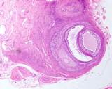
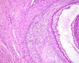
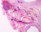
There are 54 cases in the YB database. Some contained otherwise normal ovary in fatty tissue, some had suture material adjacent to ovarian tissue, some had an attached uterine tube, one had uterus as well, 2 had ovarian neoplasia and one was in the omentum. identifying actual ovarian cortical tissue is made easier by the presence typical ovarian structures such as follicles, corpora lutea, or intersitial endocrine cells for which there are examples below.

Figure : Ovarian remnant with cystic follicles and corpora lutea

Figure : Ovarian remnant. Hydrosalpynsx and small ovarian remnant (lower right).

Figure : Ovarian remnant with follicles and interstitial cells

Figure : Ovarian remnant with follicles and interstitial cells.

Figure : Ovarian remnant with suture material (central) ovarian tissue (upper left) and dilated rete ovarii (lower right).

Fritsch DA, Allen TA, Dodd CE, Jewell DE, Sixby KA, Leventhal PS, Brejda J, and Hahn KA (2010) A multicenter study of the effect of dietary supplementation with fish oil omega-3 fatty acids on carprofen dosage in dogs with osteoarthritis. J Amer Vet Med Assoc 2010, 236: 535-539.

Heffelfinger DJ. (2006) Ovarian remnant in a 2-year-old queen. Can Vet J. 47(2):165-167.

Miller DM (1995) Ovarian remnant syndrome in dogs and cats: 46 cases (1988-1992).

Wallace MS. (1991) The ovarian remnant syndrome in the bitch and queen. Vet Clin North Am Small Anim Pract. 21(3):501-507
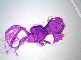
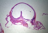
Colonic stricture

There are several reports where cats developed a fibrous ring on the serosa of the colon that lead to colonic obstruction. The cause of the ring was not determined, but local trauma, inflammation of the uterine stump and local contamination were considered. (Coolman et al 1999, Remedios and Fowler 1992, ).

I had access to one such case.


Coolman BR, Marretta SM, Dudley MB, Averill SM. (1999) Partial colonic obstruction following ovariohysterectomy: a report of three cases. J Am Anim Hosp Assoc 35(2):169-172.

*Muir P, Goldsmid SE, Bellenger CR. (1991) Megacolon in a cat following ovariohysterectomy. Vet Rec 129(23):512-513

Remedios AM, Fowler JD (1992) Colonic stricture after ovariohysterectomy in two cats. Can Vet J 33: 334-336.

*Smith MC, Davies NL. (1996) Obstipation following ovariohysterectomy in a cat. Vet Rec. 138(7):163.

Stump pyometra

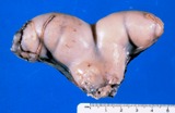
Inflammation and other disease of the uterine stump is much less common than ovarian remnants. There are 4 cases in the YB database. Two had suppurative inflammation, including a reaction to suture material, one had cystic endometrial hyperplasia and one was a normal stump. Surgery is performed on the stump when there is a discharge (especially bloody) from the vulva, or as part of surgery for an ovarian remnant (1 case had an ovarian remant).

Figure : Stump pyometra in a cat.

Photo compliments of Department of Pathobiology, OVC, Univ of Guelph


Hemorrhage/hemoabdomen

Hemorrhage and hemoabdomen is rare and the surgical pathologist may be called upon to exclude other complicating factors, such as underlying disease.


Hydronephrosis

It is indeed unfortunate when surgery for neutering or for an ovarian remnant results in ligation of a ureter. This is fortunately rare.

Perioperative mortality


Levya et al (2017) reported on perioperative mortality in dogs and cats. They spayed 71,557 cats and 47,349 dogs. 34 cats and 4 dogs died in the perioperative period (within 24 hours of preoperative sedation). The majority were in the postoperative period with those in the preoperative period second in number.

Levya JK, Bardb KM, Tuckera SJ, Diskanta PD, Dingmana PA. Perioperative mortality in cats and dogs undergoing spay or castration at a high-volume clinic. The Vet J 2017; 224: 11-15

Uterovaginal fistula

Allen and Webbon (1980) report 2 cases of vaginoureteral fistulae. These were assumed to be acquired after ovariohysterectomy as the incontinence only began after surgery.


Allen WE, Webbon PM. (1980) Two cases of urinary incontinence in cats associated with acquired vagino-ureteral fistula. J Small Anim Pract. 21(7): 367-371.

Small intestinal incarceration.