Failure of Pregnancy in Sheep

General approach.

Please refer to the general discussion of Failure of Pregnancy for basic concepts. The following is focused on ovine failure of pregnancy.

A problem of failure of pregnancy is established when the number of pregnancies that fail is above the normal for the flock or is above a level attainable for that flock. The 'normal' or anticipated rate for abortion in a large number of sheep varies from 1 to 5% and for stillbirth to be from .7 to 6%.

Surveys of reproductive loss have variable proportions of those that die from abortion, stillbirth or perinatal mortality.

Large studies suggest that abortion, stillbirth and perinatal mortality represents about 17, 20, and 63% respectively, of the loss.

Once a problem is recognised and verified, the approach taken will vary with the definition of the problem, and on the flock history.

The causes of failure of pregnancy are usually divided into two main groups - those with lesions and a recognisable cause (often nfectious) and the others. Infectious causes are usually the easiest to recognise because the lesions in the placenta can be recognised.

Based on submitted materials, from 48 to 56% of cases in sheep go undiagnosed by laboratories.

The table below provides information on diagnoses from two laboratories. Notice they have a category that is 'noninfectious'. This group refers to causes of FOP for which as cause is recognised but which is not infectious - goitre from iodine deficiency, congential anomalies and other diseases are included here.

Cause Dakota AHL Netherlands
No diagnosis 56 45 26
Noninfectious 5 2 5
Infectious 39 55 61
Placentitis 25 9 11
Campylobacter 10 12 20
Chlamydia 5 11 13
Toxoplasma 11 19 15
Coxiella 0.1 7 0

Other important causes

Pestivirus D (Border disease virus)
Brucella ovis
Listeria monocytogenes
Wesselbron virus
Rift Valley fever phlebovirus
Schmallenburg orthobunyavirus
Cache Valley orthobunyavirus
Salmonella enterica subspecies enteric serovar abortusovis
Iodine deficiency


If a known cause can be determined, a plan can be devised to control the disease, and it is much easier to provide information on the likely outcome and time to recovery. Information on this will be provided below.

There is considerable variation in the causes of failure of pregnancy from one location to another. In some places, campylobacteriosis is the most common, in others it is toxoplasmosis, or Akabane virus. From a Canadian and Ontario context, the classic causes are listed in Table 1 above.

Diagnostic process

Take precautions

It does not take long after reading the list of potential infectious causes of pregnancy failure to realise that the major infectious agents are potentially zoonotic. Of the top four causes, Coxiella burnetti is the most infectious and is well-known for causing community outbreaks. The spores of this agent are highly infectious and are spread by aerosol. They are long-lived and present in contaminated surroundings including dust in barns. sterilisation or rendering of spores in active is extremely difficult. Of the other agents, Campylobacter is spread by feco-oral transmission and Chlamydia is spread a variety of ways also. The precautions taken should reflect the potential risk. Even a closed flock that is known to be negative for Coxiella could have a breakdown in infection control. Diagnostic laboratories who handle potentially infectious material on a regular basis have standard operating procedures that include the use of special biohazard chambers, and the use of personal protective equipment that not only includes coveralls and gloves, but the special respirators and masks that filter ultrafine particles. An N95 mask with appropriate and verified fit testing is routinely used. In a field situation, use of suitable personal protective equipment including a system with a respiratory filter is highly recommended, particularly in those locations where vaccination for Coxiella is unavailable. Because many of the infectious agents are particularly hazardous in pregnancy, those individuals who are pregnant and who are in an altered immune competent state including the very young and the old are strongly advised to have no potential contact with these infectious agents. Appropriate disinfection, sanitisation and personal hygiene is a very important component to minimising risk of infection. Obviously the same recommendations hold for producers, farmers and their families or visitors.

Selection of material to examine

The general recommendations for selection of material and the numbers of cases to examine vary with the particular situation including the economic benefit. For individual cases, it is best to submit all placental and fetal tissues to an appropriate diagnostic laboratory for evaluation. It is best to provide the diagnostic laboratory with as much suitable and relevant information as possible, and to provide a maximum dollar figure for the investigation.Tentative diagnoses can be made by examining the placenta and fetus and will be described below. If there is an outbreak, a so-called abortion 'storm', a representative sample should be examined and if appropriate, submitted to the laboratory. At the field level, as many placentas and fetuses as possible should be examined and representative samples submitted for further diagnostics, if appropriate.

It is critical to submit the whole placenta, or at least a sample of a placenta that contains cotyledon and intercotyledonary placenta, and particularly any lesions.

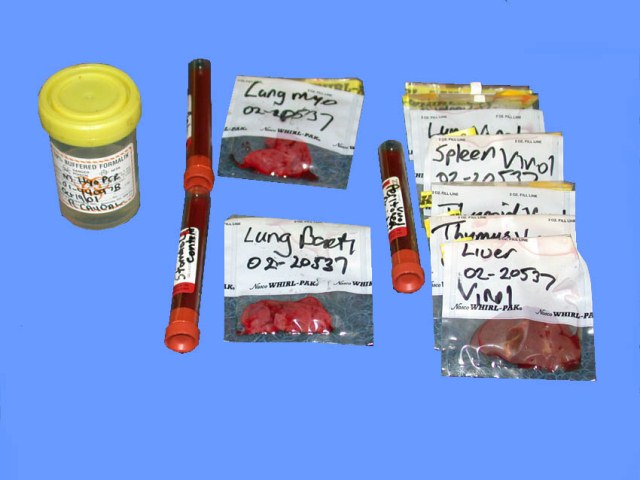
The best samples to send to a diagnostic laboratory are whole placentas and foetuses. If their transportation is not possible, selection of samples should be done in accordance with the recommendations of the diagnostic laboratory. All diagnostic laboratories provide basic information on the samples they require and these instructions should be followed to the letter. A typical general submission requires a large number of samples that are appropriately taken, labelled, and packaged.

Generalsamples

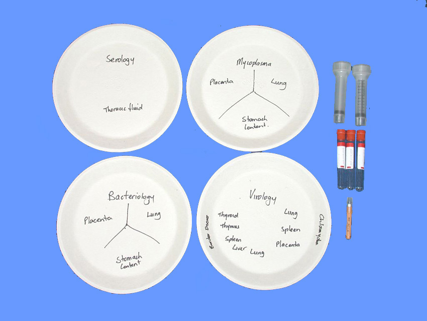
Typical containers appropriately labelled as per Users Guide of Animal Health Laboratory, University of Guelph.


Examination of Ewe(s)

Some infectious diseases are not easily confirmed - either because the agent is is very difficult to culture or identify, affects the fetus earlier in gestation and is no longer present, is neutralised by antibodies or myriad other causes. Serology may be the most accurate way of identifying the influence of an infectious agent. Collecting serum samples from a representative sample of the flock including serum from ewes with failure of pregnancy, non-pregnant and unaffected pregnant ewes should provide additional confirming evidence. It is best to check with the diagnostic laboratory if this option is available.

It is rare for reproductive samples to be taken from ewes however as a general principle, sampling of endometrial fluids may be an alternative to sampling a placenta if the placenta is no longer available

Examination of lamb(s)

Examination of a lamb or lambs is a much more involved process of examining a ruminant placenta.a thorough examination involves making observations that are different to those normally made at necropsy. In particular, recording the approximate age or length of gestation at the failure of pregnancy, identifying Fetal autolysis, and selectively examining particular tissues to achieve maximum diagnostic potential are required.

Age of lamb at loss

Most of the failures of pregnancy in sheep occurs late in gestation, and many people do not measure specific information on the degree of fetal development. Richardson et al (1976) provides information of the aging of fetal lambs. The classic measure is crown - rump length (they called it crown to anus length). This was accurate between 50 and 100 days of gestation. it is also worthwhile noting degree of wool cover and the type of coat. These measures should be written on any submission sheets and they form part of the record of your investigation

Time since death en utero

Many people are unaware of the normal changes of foetal autolysis and much of the older and even some of the newer literature lists autolytic changes as part of the spectrum of lesions of disease. Although this is not a critical observation in sheep, it is still worthwhile, conceptually, to determine if the fetus initiated its own birth (no autolysis) or whether it died rapidly en utero and became autolysed. In general, autolysis in stillborns is minimal, but autolysis in foetuses progresses rapidly because they are held at body temperature. Fetal autolysis is very different to autolysis of adult animals because they are in a sterile environment and do not have any intestinal flora.

Dillman and Dennis (1976) provided a detailed account of the autolytic changes that occur in ovine foetuses. A brief synopsis of their findings is provided in the following table.

Time since death

Change

12 hours

cornea cloudy, amnionic fluid blood tinged

24 hours

fluid in body cavities

36 hours

gelatinous fluid in subcutis

72 hours

Eyes dehydrated

144 hours

carcase dehydrated, no abomasal contents


Dillman RC, Dennis SM. Sequential sterile autolysis in the ovine fetus: macroscopic changes. Am J Vet Res. 1976; 37: 403-407.

Collection of samples

The post mortem examination of the fetus is very similar to that of an adult, at least in the initial phases. All necropsy is should be "problem based" and in the case of a fetal necropsy the aim is to identify those lesions that are significant to failure of pregnancy and to concentrate on those organs and tissues which are likely to have lesions. The following is a list of organs that should be sampled in every case – some of these are rarely sampled in an adult. A list of samples to take is usually provided by the diagnostic laboratory and to ensure all samples are taken the list of samples can be written on a paper plate and as the necropsy proceeds, pieces of tissue can be placed on the appropriate sample name on the plate.

Gensamplcollect

Organs to be sampled in every fetus, in addition to routine samples from all major organs:

  1. Brain
  2. Thyroid gland
  3. Abomasal content
  4. Femur


Dillman RC, Dennis SM (1976) Sequential sterile autolysis in the ovine fetus: macroscopic changes. Amer J Vet Res 1976, 37: 403-407.

Examination of placenta(s)

Measurement of placental and fetal weight is a routine procedure for many species. Fetal weight is well known to be related to perinatal mortality with a birth weight above 5kg being a major risk factor for dystocia, stillbirth and subsequent mortality in the first hours of life.

Placental weight is seldom measured in sheep, except in a research setting. It is known that placenta weight increases above normal in midgestation (between 30 and 96 days) if there is maternal undernutrition. This is presumably a mechanism to compensate as fetal growth is not affected by undernutrition during this stage (McCrabb et al 1991).

Placental lesions in infectious disease can contain huge numbers of highly infectious particles and it is essential to take precautions (see above).

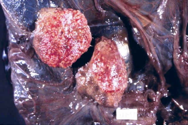
Lesions fall into two main types - cotyledonary necrosis and intercotyledonary placentitis. The lesions of focal cotyledonary necrosis are subtle and are often missed. Placentitis is usually very obvious.

Focal cotyledonary necrosis is the hallmark lesion of Toxoplasmosis. All cotyledons are usually affected and in most cases about 50% of the cotyledonary area. There are 1or 2mm white foci throughout the cotyledons.

Toxo1

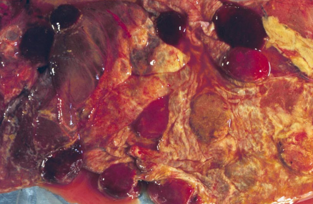
Placentitis comes in 2 forms with the chronic form being the most common. The acute form is more difficult to identify. The classical chronic changes are

Chlamydia02

Chlamydia

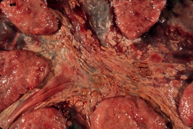
The typical acute lesions are subtle:


Lesions present

Because in sheep the most common location for lesions is the placenta, the most recognised of causes is placental disease. It is very important that any investigation includes examination of the placenta. Placentitis and multifocal placental cotyledonary necrosis are the main lesions.

Fetal lesions occur periodically. Iodine deficiency and the resultant goitre is a regional disease. Congenital anomalies are uncommon and usually sporadic.

Infectious

It is preferable to correlate infections and lesions to establish causality. The sophisticated testing now possible will identify infectious agents in many placentas but identifying their role in the failure of pregnancy is more difficult. Hazlett et al (2013) found evidence of infectious agents - Coxiella burnetti in 113 (69%) of 163 placentas of aborted lambs, Chlamydia abortus in 42 (26%) of 162 placentas, and only a small percentage of these placentas had sufficient lesions to attribute the infectious agent to the cause of FOP (10% for Coxiella and 38% for Chlamydia). Based on this study, coinfection is common, and all 4 common agents (Chlamydia, Coxiella, Campylobacter and Toxoplasma) can occur in various combinations in each placenta.

Clune et al (2021) Reported on 529 investigations of failure of pregnancy – abortion and stillbirth – in Australia. They found an infectious cause in 46% of investigations. In an additional 30% of cases, there was evidence of an infectious cause such as placentitis, hepatitis or fetal pneumonia and found that in the infectious causes Campylobacter species represented 32.3%, Listeria was 25.7% and Toxoplasma gondii was 9.3%. Other agents represented 14% of cases.

Hazlett MJ, McDowall R, DeLay J, Stalker M, McEwen B, van Dreumel T, Spinato M, Binnington B, Slavic D, Carman S, Cai HY. A prospective study of sheep and goat abortion using real-time polymerase chain reaction and cut point estimation shows Coxiella burnetii and Chlamydophila abortus infection concurrently with other major pathogens. J Vet Diagn Invest 2013; 25: 359-368

Diagnosis

Borel et al (2014) provide a review of major causes of abortion in ruminants in Europe. These include viral, bacterial and fungal causes listed below.

Correlation of recovery of infectious organisms and histological lesions is the hallmark of diagnosis. Molecular methods such as PCR and qPCR are all the rage.

Gutierrez et al (2014) found that rtPCR was an valuable addition to routine diagnosis. They performed qPCR and correlated it with lesions to identify the level of organism RNA that could be associated with disease.


Borel N, Frey CF, Gottstein B, Hilbe M, Pospischil A, Franzoso FD, Waldvogel A. (2014) Laboratory diagnosis of ruminant abortion in Europe. Vet J 2014; 200: 218-229.


Gutierrez J, O'Donovan J, Proctor A, Brady C, Marques PX, Worrall S, Nally JE, McElroy M, Bassett H, Fagan J, Maley S, Buxton D, Sammin D, Markey BK. Application of quantitative real-time polymerase chain reaction for the diagnosis of toxoplasmosis and enzootic abortion of ewes. J Vet Diagn Invest. 2012; 24: 846-854.

Bacteria

General considerations

The exact cause of failure of pregnancy in bacterial disease is debated. In endotoxin producing infections there is the production of a fetal acute systemic inflammation. Endotoxin in amniotic fluid and in the lung will induce this response.

Kemp MW, Kannan PS, Saito M, Newnham JP, Cox T, Jobe AH, Kramer BW, Kallapur SG. Selective exposure of the fetal lung and skin/amnion (but not gastro-intestinal tract) to LPS elicits acute systemic inflammation in fetal sheep. 2013; 8: e63355

The big four infectious causes of infectious abortion in sheep are Coxiella burnetti, Chlamydia abortus, Campylobacter sp, and Toxoplasma gondii. Each will be discussed in detail below.

Family Brucellaceae, Genus Brucella

Brucella ovis

Ovine brucellosis is rare in the eastern United States and Canada.  It is an important cause of disease in rams in a both Western US and Western Canada. The primary presenting condition is epididymitis of rams. In most flocks, abortion occurs only occasionally. Infection of ewes is usually by infected rams at mating and the infection is cleared spontaneously.  It is possible for outbreaks to occur and to have an abortion storm of up to 70%.

Introduction of infection into a flock usually occurs with the introduction of an infected ram. Control of the disease is the best achieved by elimination of infected rams using scrotal palpation and serology outside of the breeding season. In flocks that are heavily infected, vaccination may be helpful but it is only available in the USA. It is important to prevent infection and this requires purchasing rams only from flocks known to be Brucella free.

Family Campylobacteraceae, Genus Campylobacter

Campylobacter fetus, Campylobacter jejuni

Abortion due to Campylobacter fetus subsp fetus and subsp intestinalis, and Campylobacter jejuni causes an intercotyledonary placentitis with abortion and stillbirths, and the birth of weak lambs.(Campylobacter fetus subsp venerealis is a reproductive pathogen of cattle). Ewes are infected through the feco-oral route and carriers have the bacterium within the gall bladder. Aborted fetuses and placentae are a source of infection, but outbreaks are not as dramatic as with chlamydiosis. The incubation is 7 to 60 days, and abortion can ‘cycle’ within a lambing season.

Gross and microscopic lesions can be either "acute" placentitis as defined above or they may be chronic.

Yaeger et al (2021) reported on the changes of Campylobacter abortion (including jejuni, fetus fetus and unidentified species). Macroscopic lesions of placentitis were seen in 6% of 120 cases and hepatic lesions were seen in 4%. Microscopic lesions were seen in 93% of placentas, with bacterial colonies in chorionic vili (stroma) in 54%. Colonies in trophoblasts were much less common than colonies in the stroma or subtrophoblastic blood vessels. Vasculitis was seen in only 13%. In other organs, there was suppurative fetal pneumonia in 48%, necrosuppurative hepatitis in 16%, and suppurative meningitis in 7%.


Yaeger MJ, Sahin O, Plummer PJ, Wu Z, Stasko JA, Zhang Q. The pathology of natural and experimentally induced Campylobacter jejuni abortion in sheep. J Vet Diagn Invest. 2021 Nov;33(6):1096-1105. doi: 10.1177/10406387211033293. Epub 2021 Jul 26. PMID: 34311616; PMCID: PMC8546477.


Family Helicobacteraceae, Genus Helicobacter

Helicobacter

Lesions identical to that of Campylobacter fetus is seen with sporadic infection with Helicobacter species ('Flexispira' rappini taxa). H bilis and H trogontum are bacteria identified in field cases.

Gill J, Haydon TG, Rawdon TG, McFadden AM, Ha HJ, Shen Z, Feng Y, Pang J, Swennes AG, Paster BJ, Dewhirst FE, Fox JG, Spence RP. Helicobacter bilis and Helicobacter trogontum: infectious causes of abortion in sheep. J Vet Diagn Invest. 2016; 28: 225-234.

Family Chlamydiacae, Genus Chlamydia

Chlamydia abortus

Chlamydophila as a genus name did not last - it is now called Chlamydia again.

Chlamydia abortus is very important in sheep, both as an infectious agent and as a potential zoonosis. Chlamydial abortion is also called enzootic ovine abortion, and enzootic abortion of ewes, and the lesion is chronic placentitis.

Ewes are infected through mucous membranes including those of the mouth, conjunctiva and reproductive tract. The source of the agent is aborted fetuses and uterine discharge, vaginal secretions of carrier ewes at estrus or there is spread through the prepuce and semen of rams (at least temporarily). After exposure to Chlamydia abortus, sheep develop antibody, but infection can be detected for a month or more as interstitial pneumonia or focal hepatitis. The organism can be found in the mononuclear cells of the uterus. From there they infect the epithelial cells of the placentomes, and control of infection is by neutrophilic infiltrates. Trophoblasts in the periplacentomal region become infected and there is a logarithmic increase in numbers, with subsequent necrosis and inflammation of the placenta. Maternal inflammation becomes restricted to lymphocytic inflammation around the endometrial cells of the uterine glands. It is assumed that this immune reaction prevents infection of the placenta in subsequent pregnancies.

The incubation period during gestation is very long -- from 50 to 90 days. If a ewe is infected early in gestation, she will abort in the same gestation (gestation is 145 days). If infection occurs in late gestation she will abort during the next pregnancy. In general, once a ewe aborts, she will not abort a second time.  They may remain as carriers for several years however. Exactly why and how the immune reaction to Chlamydia develops and interacts in the pregnant uterus is being unravelled (slowly!)

Introduction of infected replacements is a common way to introduce infection into a naive flock and to begin an abortion storm. In the first year following introduction there may be a few animals abort but most of the flock will become infected. Abortions normally occur the following year with up to 75% of pregnant ewes aborting. In subsequent years the disease becomes enzootic and only first time lambers abort.

Diagnosis of infection can be made on aborted tissues especially the placenta, and flock serology is possible in some countries. There is a low level of false positives and there is some cross reaction with Chlamydia pecorum, a Chlamydia found the faeces.

Treatment in the face of an outbreak of Chlamydia abortion includes the use of long acting oxytetracycline. This should be repeated every 10 to 14 days. Orally administered tetracycline is an alternative. Because of the long incubation period, there is usually a poor to moderate efficacy of this treatment.

Vaccination is possible with a killed vaccine were available and has a variable efficacy.

Chlamydia pecorum

There are reports of Chlamydia pecorum causing infertility in sheep, including abortion.


Borel N, Polkinghorne A, Pospischil A. A Review on Chlamydial Diseases in Animals: Still a Challenge for Pathologists? A Review on Chlamydial Diseases in Animals: Still a Challenge for Pathologists? Vet Pathol. 2018; 55: 374-390.

Walker E, Lee EJ, Timms P, Polkinghorne A. Chlamydia pecorum infections in sheep and cattle: A common and under-recognised infectious disease with significant impact on animal health. Vet J. 2015; 206: 252-260.

Family Coxiellaceae Genus Coxiella

Coxiella burnetii

Coxiellosis is a recognised cause of failure of pregnancy in sheep, but overall, it is less important than in goats, where it is common. Some sheep flocks are severely affected and in others it is a sporadic cause. Prevention of its occurrence in a flock is very important both from a management and public health point of view - Coxiella burnetii is extremely infections and one 'spore' is the infectious dose.

The herd history of coxiellosis is very similar to that of Chlamydia. Adults show no clinical disease, but the organism grows to large numbers in the pregnant uterus where

The pathogenesis of Q fever begins with the organism. It is an obligate intracellular gram-negative bacterium that is closely related to Legionella, Francisella and rickettsias. It is highly resistant to physical and chemical degradation, and is infective in very low doses – as low as one infectious particle – making it the most infectious agent known. Aerosolisation as ‘dust’ is significant. Naïve animals are infected by aerosol or from infected unpasteurised milk. After primary replication in local lymph nodes, it becomes bacteraemic and localizes in the mammary gland and pregnant uterus. It can be found in large numbers in amniotic fluid and in the placental membranes, especially trophoblasts. Some animals become carriers and shed the organism in milk and in uterine fluids. Shedding in the periparturient period is especially common.

While goats can be persistently infected and shed the organism at subsequent kidding, . Because the organism can be highly ineffective in a dried state, it is dust born, and barns and sheds become contaminated and can be potentially infective for many years. Infection of the premises can also occur with carrier animals including sheep, cattle, cats, birds and other wildlife.
Treatment in the face of an abortion storm can be achieved through the use of oxytetracycline as with chlamydiosis. Special precautions are required for any infective environment and when assisting kidding. The use of gloves, appropriate masks and eye protection while assisting in kidding or cleaning barns is strongly advised. Children, elderly and pregnant individuals should stay out of the barn.


Family Helicobacteraceae, Genus Helicobacter

Helicobacter bilis; Helicobacter trogontum (previously Flexispira rappini Taxon 1 through 10)

The organism previously known as Flexispira rappini or "F. rappini" taxa (1 through 10) are now considered to be helicobacters and either Helicobacter bilis or Helicobacter trogontum based on 16S and 23S ribosomal RNA sequences. Flexispira rappini was considered a cause of sporadic infection and did not cause outbreaks. The lesions are identical to those of campylobacteriosis.

Gill et al (2016) reported on extensive work in identifying Helicobacter bilis and Helicobacter trogontum in abortion in New Zealand. Most submission of aborted lambs did not have placenta. The lambs were fresh and no autolysis. The affected lambs had round pale up to 10mm foci in the liver capsule. Within the liver the foci were up to 30mm diameter. These were, histologically, regions of necrosis and thrombosis of centra veins. Organisms are visible at the edge of the liver lesions. Some lambs had portal hepatitis too. Bacteria were visible with silver stain (they used Warthin Starry).

The organism is present in the intestine of sheep, and dogs, so it is likely that helicobacters are spread from dogs to sheep, and then after intestinal infection, portal spread of the organism to the liver and bacteremia localises in the pregnant uterus leading to placentitis. Intestinal intake of bacteria in the fetus would then result in liver lesions and bacteremia.


Gill J, Haydon TG, Rawdon TG, McFadden AM, Ha HJ, Shen Z, Feng Y, Pang J, Swennes AG, Paster BJ, Dewhirst FE, Fox JG, Spence RP. Helicobacter bilis and Helicobacter trogontum: infectious causes of abortion in sheep. J Vet Diagn Invest. 2016; 28: 225-234.


Family Listeriaceae genus Listeria


Listeria monocytogenes

Family Leptospiraceae genus Leptospira

Leptospira interrogans serovars Hardjo, Bratislava, Ballum and Pomona

Sheep do not commonly develop leptospirosis but can be infected by consuming water contaminated with leptospires from other species. The last 2 weeks of gestation and first week postpartum are the main times when lambs will be affected. Stillborn lambs and weak lambs may have lesions

Family Salmonellaceae; Genus Salmonella

Salmonella enterica subsp enterica serovar Abortusovis


Salmonella enterica subsp enterica serovar Brandenburg


Protozoa

Neospora caninum

Many animal species are susceptible to infection with Neospora caninum and some, especially cattle, develop failure of pregnancy and perinatal mortality. Experimental infection of sheep with this protozoa resulted in seroconversion and abortion, but only in those infected at days 65 and 90. Those infected at day 120 of gestation had normal lambs. Most of those that aborted (46 of 51) had brain lesions of multifocal necosis, mineralisation and gliosis. There are isolated reports of natural cases of failure of pregnancy with this organism, but it does not appear to be common.

Toxoplasma gondii

Toxoplasmosis can play a major role in ovine abortion.

Macroscopic lesions

The lesions are grossly visible focal necrosis in the cotyledon and histologic changes in the brain of fetuses. The cotyledonary changes are very characteristic and are those of multifocal necrosis restricted to the cotyledons. There are no macroscopic lesions in the brain of affected fetuses.

Toxo1

Microscopic lesions

The microscopic lesions are the histological correlates of focal necrosis seen grossly. Necrosis is multifocal, randomly distributed in the cotyledon although in many cases be focal necrosis appears to be fairly regularly distributed. The placenta is sometimes mummified and histological identification of the areas of necrosis requires a keen eye on low magnification. Immunohistochemical identification of Toxoplasma antigen is particularly useful in identifying the agent as the tachyzoites are virtually impossible to see and the cyst forms can be very infrequent at best.sometimes tacky so-ites are visible within trophoblasts.

Histological lesions in the fetus can be variable also. Some report only the presence of a leucoencephalomalacia and in about 20 to 30% of cases, presumably the result of fetal hypoxia. Some report the presence of a typical focal gliosis and necrosis, and organisms in the tissue but this does not appear to be very common. A report on experimental infection indicates that lesions are most commonly found in the optic tract, the rostral pons and the 4 mm caudal to the ansate sulcus. These locations are not usually sampled in routine examination of the brain. There are isolated reports of lesions, including cysts, occurring in the myocardium.

Toxoplasmosis in humans is associated with congenital defects including hydrocephalus, microcephalus and mental retardation. There are only isolated reports of sheep with such lesions.

Pathogenesis

Infection with Toxoplasma is widespread and serological surveys suggest that 65 to 95% of a flock will have evidence of previous infection. The source of infection is usually infected cats which pass oocysts in their faeces for approximately seven days. Oocysts can remain infective for six months. Naive ewes will develop a protective immunity but if they are pregnant, infection of the cotyledon will result in abortion.

The earlier in gestation infection occurs, the more necrosis and less inflammation occurs. In the fetus, brain lesions are necrosis early in gestation, while focal gliosis occurs more commonly in late gestation. In some animals there is vertical tranmission of organisms to the fetus, Others have the more classical abortion profile.

Classical abortion with Toxoplasma gondii occurs about 28 days after exposure. There can be an 'acute' presentation with abortion in 7-14 days after exposure, and experimentally this is induced with exposure to large numbers of oocyts.


Control of toxoplasmosis involves removing the source of oocysts. Control of rodents and cats and their access to feed and pasture is particularly important. Even this will not guarantee complete control as purchased feed may be contaminated. Feral cats are very difficult to control also!

Prevention of infection or abortion can be achieved by including monensin or decoquinate in the feed throughout gestation. A vaccine is available in some countries.


Benavides J, Fernández M, Castaño P, Ferreras MC, Ortega-Mora L, Pérez V. Ovine Toxoplasmosis: A New Look at its Pathogenesis. J Comp Pathol. 2017; 157: 34-38.

Sarcocystis tenella

Agerholm and Dubey (2014) report on a stillborn lamb with lesions of sarcocystosis, likely Sarcocystis tenella. The lesions were in the brain and cotyledons of the placenta. They are similar to toxoplasmosis in having focal areas of necrosis surrounded by glial cells within the brain and shizonts within endothelial cells. The lesions in the cotyledons were focal areas of necrosis and mineralization.


Agerholm J, Dubey J. (2014) Sarcocystosis in a stillborn lamb. Reprod Domest Anim. 2014; 49: e60-e63


Viruses

Family Flaviviridae, Genus Pestivirus

Species: Pestivirus D (Border disease virus)

Border disease was first reported from the border region of England and Wales. In other countries like Australia, affected newborn lambs are called 'hairy shakers'. It is caused by Pestivirus D (Border Disease virus [BDV]) of the family Flaviviridae, genus Pestivirus that is closely related to Pestivirus A and B (Bovine Viral Diarrhea virus [BVDV]). The type of disease seen in lambs is similar to calves infected en utero with BVDV. Infection of a pregnant ewe can result in fetal loss, birth of weak lambs, or birth of lambs that are immune compromised. Infection at less than 65 days of gestation results in early embryonic death. Infection at 65 to 85 days of gestation can result in the birth of a persistently infected lamb that may be weak, have a hairy coat and shake (thus the clinical manifestation of a "hairy shaker"). Ewes infected after 85 days of gestation will give birth to normal lambs with titres to Border Disease virus.

The control of border disease revolves around reducing exposure of naive individuals during pregnancy. Persistently infected sheep provide a source of infection so identifying and removing them from the flock is essential. Vaccination of ewes with a killed BVD vaccine is not effective as there is no cross protection. Border disease is a self-limiting disease if persistently infected animals are identified and removed. They can be identified by virus isolation from the blood (buffy coat) of infected animals.

Brain lesions in infected lambs include lymphocytic and necrotic meningoencephalitis and hypomyelinogenesis in the periventricular regions, cerebellum and brain stem. Leukomalacia can be seen also. Cerebellar atrophy can also occur (Toplu et al 2011). Hypomyelinogenesis is probably because of lower than normal thyroxine (T4) and T3 concentration from viral effects on the thyroid. T3 and T4 are required to allow oligodendrocytes to produce myelin normally.

Hairyness is likely a direct effect of the virus on hair follicles.

BDV infects vessels and endothelium of caruncles and produces endometritis and focal to confluent necrosis of the caruncle.

Species: Pestivirus A, B (Bovine viral diarrhea virus)

Bovine viral diarrhea virus infection of sheep produces an identical range of lesions as BDV, which is a closely related virus. It results in embryonic loss, abortion, stillbirth and birth of lambs with malformations as expected with central nervous affects of the pestivirus. Contamination of an orf vaccine with Pestivirus B caused an outbreak in Spain in 2017. Neurological lesions include hydranencephaly and cerebellar hypoplasia. White matter of the spinal cord had rarification, spongiosis and hypomyelination.


Asín J, Hilbe M, de Miguel R, Rodríguez-Largo A, Lanau A, Akerman A, Stalder H, Schweizer M, Luján L. An outbreak of abortions, stillbirths and malformations in a Spanish sheep flock associated with a bovine viral diarrhoea virus 2-contaminated orf vaccine. Transbound Emerg Dis. 2021; 68: 233-239.

Elvira Partida L, Fernandez M, Gutierrez J, Esnal A, Benavides J, Perez V, de la Torre A, Alvarez M, Esperon F. Detection of Bovine Viral Diarrhoea Virus 2 as the Cause of Abortion Outbreaks on Commercial Sheep Flocks. Transboundry Emerg Dis 2017; 64: 19-26


Family Bunyaviridae, Genus Orthobunyavirus

Species: Akabane orthobunyzvirus, Aino orthobunyavirus, Cache Valley orthobunyavirus (CVV, aka Bunyamwera virus), Schmallenberg orthobunyavirus

Viruses of the genus Orthobunyavirus are spread by insects of the Culicoides genus. The virus species of interest in this group are Akabane orthobunyavirus and Aino orthobunyavirus ( found in the asiatic and asiopacific area), Cache Valley orthobunyavirus, found in USA and Canada), and Schmallenberg orthobunyavirus ( identified in Europe in 2011).

Immunocompetent animals develop a transient infection and become immune. Naive animals who are pregnant also develop a subclinical infection but the virus crosses the placenta and infects the fetus where its primary target is the nervous system. These viruses are referred to as neurotropic viruses and they cause necrosis of neurones and progenitor cells especially of the periventricular regions and in some individuals also induce encephalitis. The lesions in the central nervous system of fetuses that are immunocompetent result in either subclinical infection or histological changes of lymphocytic inflammation typical of a viral infection. Those fetuses that are not immune competent may develop hydranencephaly, porencephaly, cerebellar hypoplasia, and micromyelia with the subsequent development of arthrogryposis, muscle hypoplasia and a variety of musculoskeletal malformations.

Cache Valley orthobunyavirus

Infection of the fetus between 28 and 48 days of gestation can result in fetal death or musculoskeletal and central nervous system malformations.The virus is eliminated within several weeks. If the fetus goes to term, the virus cannot be recovered from fetal tissue. Fetal immunocompetence is at 70 to 75 days.

The macroscopic lesions include oligohydramnios, scoliosis, torticollis, and arthrogryposis. Fetal mummification can also occur.

Microscopically, there is severe multifocal to extensive necrosis in the cerebral cortex, spinal cord grey matter and especially dorsal horn, and skeletal muscle. Focal lymphocyte aggregates may be present in the brain and meninges, and in the skeletal muscle. Virus is present within the areas of necrosis and in neurones. Viruses also found in my sights around the spinal column.

Waddell et al (2019) wrote a scoping review of CVV infection.


Rodrigues Hoffmann A, Dorniak P, Filant J, Dunlap KA, Bazer FW, de la Concha-Bermejillo A, Welsh CJ, Varner P, Edwards JF. (2013) Ovine fetal immune response to Cache Valley virus infection. J Virol. 2013; 87: 5586-5592.

Rodrigues Hoffmann A, Welsh CJ, Wilcox Varner P, de la Concha-Bermejillo A, Marchand Ball J, Ambrus A, Edwards JF.(2013) Identification of the target cells and sequence of infection during experimental infection of ovine fetuses with Cache Valley virus. J Virol 2012; 86: 4793-4800.

Waddell L, Pachal N, Mascarenhas M, Greig J, Harding S, Young I, Wilhelm B. Cache Valley virus: A scoping review of the global evidence. Zoonoses Public Health. 2019; 66: 739-758.


Schmallenberg orthobunyavirus

In a study of 40 ovine cases, 37 aborted foetuses had arthrogryposis, 14 had brachygnathia inferior, and 15 had curvature of the spine either torticollis, kyphosis, lordosis or scoliosis. Lesions in the brain included unilateral or bilateral internal hydrocephalus in 26 (diagnosed as dilation of the ventricles) and six cases of hydranencephaly (identified as segmental or complete loss of the brain without discernible grey and white matter and nearly complete loss of the cerebral cortex. 26 cases had cerebellar hypoplasia.
Histologically there was perivascular cuffing with lymphocytes and histiocytes, glial nodules in the mesencephalon and hippocampus in 10 ovine cases. There was also necrosis of the neurophil with accumulation of get a cells and vacuolar lies a and mineral deposition. Cerebellar hypoplasia was identified as a reduction in the number of Purkinje neurones. There was also demyelination and diffuse astrogliosis and all microgliosis.

Hypoplasia of skeletal muscle is the main non-nervous system change (Seehusen et al 2014). Viral antigen nor RNA is detectable in the peripheral organs.


Herder V, Wohlsein P, Peters M, Hansmann F, Baumgärtner W. (2012) Salient lesions in domestic ruminants infected with the emerging so-called Schmallenberg virus in Germany. Vet Pathol 2012; 49: 588-591.

Peperkamp NH, Luttikholt SJ, Dijkman R, Vos JH, Junker K, Greijdanus S, Roumen MP, van Garderen E, Meertens N, van Maanen C, Lievaart K, van Wuyckhuise L, Wouda W. Ovine and Bovine Congenital Abnormalities Associated With Intrauterine Infection With Schmallenberg Virus. Vet Pathol. 2015; 52(6):1057-1066.

Seehusen F, Hahn K, Herder V, Weigand M, Habierski A, Gerhauser I, Wohlein P, Peters M, Varela M, Palmarini M, Baumgartner W. (2014) Skeletal muscle hypoplasia represents the only significant lesion in peripheral organs of ruminants infected with Schmallenberg virus during gestation. J Comp Path 2014; 151: 148-152.

Rift Valley fever phlebovirus

Rift Valley Fever phlebovirus is in the Order Unassigned, Family Bunyaviridae, Genus Phlebovirus

The macroscopic changes are nonspecific - petechial hemorrhage and edema.

The classical microscopic lesions of RVFV is multifocal hepatic necrosis. Odendaal reported this in 48 of 58 cases. There can be eosinophilic intranuclear inclusion bodies. There is lympholysis of lymph node and spleen, and death of trophoblasts and endothelial cells of the placenta. Antigen positive cells are in many cells of the body, not just where there is cell death. Identification of antigen and lesion is most commonly found in the cotyledon and caruncle.


Odendaal L, Davis AS, Fosgate GT, Clift SJ. Lesions and Cellular Tropism of Natural Rift Valley Fever Virus Infection in Young Lambs. Vet Pathol. 2020; 57: 66-81.

Bluetongue virus (BTV)

The bluetongue viruses are of the Order Unassigned, Family Reoviridae, Subfamily Sedoreovirinae, Genus Orbivirus

There are many viruse of the Bluetongue group. Like the Orthobunyaviruses, they are spread by insects of the Culicoides genus and they cause a similar disease to that of the Orthobunyavirus genus. Early embryonic mortality and hydranencephaly is a common manifestation of infection of pregnant ewes in early gestation.

Wesselsbron virus (WESSV)

WESSV is of the Order Unassigned, Family Flaviviridae, Genus Flavivirus and is related to yellow fever virus.

Pregnant ewes develop an initial viremia and abort with the fetus being infected. If FOP doesnt occur at this stage, the fetus becomes infected and there is single cell hepatocyte death that is mild to severe. Eosinophilic intranuclear inclusion bodies are frequent.


Fungi

Mycotic abortion is well known and recognised in cattle, but it is very rare in sheep. There are only occasional case reports.

Non infectous causes

Goitre and iodine deficiency

The Great Lakes basin and selected locales are deficient in iodine, and goitre results. Unborn lambs are particularly affected. The degree of goitre and the systemic effects on the fetus vary. Many are subclinical and the enlargement of the thyroid gland can only be detected by exposing the thyroid gland at necropsy and identifying an enlargement. The fetus may have myxedema and alopecia too, but the latter, in particular,can be difficult to identify as fetuses only develop hair late in gestation.

Biliary atresia

Occasionally aborted lambs will have prominent bile plugs or lakes. Absence of bile ductules indicates atresia. Causes include inherited disease and acquired disease. Acquired biliary atresia occurs with exposure to a plant toxin. Biliatresone is a toxin found in Dysphania plants that can induce this.


Lorent K, Gong W, Koo KA, Waisbourd-Zinman O, Karjoo S, Zhao X, Sealy I, Kettleborough RN, Stemple DL, Windsor PA, Whittaker SJ, Porter JR, Wells RG, Pack M. Identification of a plant isoflavonoid that causes biliary atresia. Sci Transl Med. 2015; 7: 286ra67

Uterine torsion

Uterine torsion can cause mummification in ewes

Jones EM, Neef A, Shearer PL, Gunn AJ. Hydroureteronephrosis and fetal mummification secondary to uterine torsion in a Merino ewe. Aust Vet J. 2020; 98: 529-532.

Congenital anomalies

Anomalies and fetal monsters attract a lot of attention. Fortunately, they are sporadic, rare and therefore of no significance from a flock perspective. The potential list of anomalies is extensive. Arthrogryposis, cyclopia, and cardiac anomalies are the more common. Arthrogryposis and anomalies of the central nervous system are particularly important, as these are also reported in outbreak situations because of viral infections such as Akabane virus in tropical countries, Schmallenberg virus in Europe and the UK, Blue Tongue virus and Cache Valley virus in the USA. Border disease virus also can produce neurological anomalies. Serological evidence of infection with Border Disease and Cache Valley viruses is occasionally found in Ontario.

Ingestion of poison hemlock (Conium maculatum) is known to produce the fetotoxic effects including flexural deformities of the carpus, but the lambs recover to be normal by eight weeks of age.

From time to time, anomalies occur in flocks and no cause can be determined.

Uterine rupture

There are sporadic reports of uterine rupture during pregnancy. One report (Mitchell 1989) indicated the rupture occurred at the site of a previous caesarian section.

No lesions present

Identifying an infectious cause of failure of pregnancy is one of the easiest steps in managing a flock with the clinical syndrome of pregnancy failure. When no lesions are present in the fetus, placenta or ewes, identifying the exact cause is much more difficult as the clues include identifying a change in management, finding a genetic link and eliminating some of the known causes. The following in a list of possibilities to be explored but for which a simple test is unavailable.

There are situations where a change in food, housing, environmental conditions, weather and many other management situations appear to be linked with failure of pregnancy. Many of these are impossible to replicate in an experimental setting to verify they are responsible.

Watson (1962) reviewed the literature on noninfectious failure of pregnancy up until that time.

Stress of Handling

There is mention of the stress of handling of ewes in late gestation as a cause of sporadic outbreaks of late term abortion. Emperical evidence is lacking.

Chromosomal defects

Research in humans and cattle suggests that early embryonic mortality is often the result of chromosomal abnormalites.

Inheritied defects

Genetists recognise that some animals alive today are heterozygotes to a particular trait. This means that monozygosity of that particular trait is lethal! These animals must die en utero or in the perinatal period. Mutations of essential genes, like that of p53, are responsible for miscarriage in humans, and there are many mutations in mice that are incompatable with survival. This defects in chromosomes, genes, gene products, translation and transcription), packaging and release are all potential targets. We currently have no way of detecting these in a diagnostic setting. We will continue to have cases of idiopathic abortion into the forseeable future.

Change in food and dietry deficiency

Malnutrition has been blamed for centuries but confirmation is difficult to achieve. Ewes rendered fed diets deficient in protein, vitamin A, and copper only had failure of pregnancy when the deficiency was extreme and the adults severely affected.

Ewes with pregnancy toxemia are known lose their lambs. Mitchel and Stratford (1987) studied the placentas of affected ewes and found a relative reduction of the caruncle as compared to the cotyledonary placenta.

Unusual diets, such as a diet of root crops are suspected in some cases. Emperical evidence is lacking.

Deficiency of copper


Deficiency of vitamin A


Mycotoxin exposure

It is well known that pregnant animals given selected mycotoxins abort. The corollary is that some instances of failure of pregnancy are the result of mycotoxin exposure. Feed and pasture can be tested for mycotoxins by diagnostic laboratories and correlations can be made.

Ergotism is thought to be involved with failure of pregnancy including stillbirth.

Ewes grazing pastures of onion grass (Romulea rosea) dominance are known to suffer failure of pregnancy and the Romula leaf spot fungus Helminthosporium biseptatum that grows on the leaves is blamed. The disease is known as 'romulosis'.

Exposure to environmental toxins

Sheep given selected drugs, toxins and toxic plant extracts abort in an experimental setting. Mycotoxin screening is a routine procedure but the screening of food and pasture samples for toxins is much more costly and difficult.

Plant toxicosis

The following are plants reported to result in failure of pregnancy, usually abortion.

Ipomoea carnea subspecies fistulosa


Embryonic mortality

Early embryonic mortality in sheep occurs in about 5 to 30% of pregnancies. Younger ewes tend to have fewer losses. Loss is high in old ewes. The tendency is for the majority of reproductive losses to occur in the first 30 days.

Boshier (1968) provides details of the histological appearance of the placental membranes of sheep at day 18 post coitus, and is a guide to identifying viable from nonviables embryos.

There is little known about the causes in sheep.

Excessive temperature

Situations where there is a high environmental temperature can affect early embryonic mortality. When temperatures are high, ewes will not eat, so an up to 30% reduction in survival of embryos could be from either higher body temperatures or from reduced food intake. Abortion or stillbirth appear not to occur.

Assisted reproductive technologies

Fertility is often reduced in assisted reproductive technologies like embryo transfer. Grazul-Bilska et al (2014) found reduced expression of mRNA of 12 of 14 angiogenic factors of placentas in embryos from assisted technologies.

Anna T Grazul-Bilska, Mary Lynn Johnson, Pawel P Borowicz, Jerzy J Bilski, Taylor Cymbaluk, Spencer Norberg, Dale A Redmer and Lawrence P Reynolds (2014) Placental development during early pregnancy in sheep: effects of embryo origin on vascularization. Reprod 2014; 147: 639-648

Stillbirth

Dystocia

About 1% of ewes normally require assistance to complete parturition. The prevalence of dystocia is up to 12% in some flocks. Normal parturition takes about 90 minutes on average. Most cases are because of oversize of the lamb and although the position of the lamb is normal, it is too big to be expelled unassisted.

Dystocia is seen when

  • Nutrition of the ewe in the latter stages of gestation is high. This is considered a major influence.
  • Some rams produce lambs with a higher birth weight and their offspring may be larger.
  • Some ewes have a smaller pelvis than those of the same breed.

Lambs with dystocia usually have subcutaneous edema of those parts that were outside the birth canal when parturition ceased. This is typically the head including the tongue and lips. Sometimes the rump and hind legs are swollen (from posterior presentation).

Lambs taking an excessive amount of time in parturition will suffer hypoxia and develop cerebral edema or simply be unable to either rise or suckle.

For more information, check the section on perinatal mortality.

Prolonged gestation

It is rare for pregnancies to go longer than the usual gestation period. There are some classic diseases where this occurs, and occasionally, outbreaks are reported.

Reports suggest that pregnancy up to 4 weeks over time. Lambs increase in weight to be up to 12 kg. Lambs usually have a central nervous system anomaly such as holoprosencephaly and cyclopia, hydranencephaly, anencephaly, and vertebral and spinal anomalies with arthrogryposis. Lambs must be caesarean delivered, or there is dystocia and maternal death. A variety of endocrine disorders occur concurrently including thyroid hypoplasia, hypoadrenocorticism with or without adrenal hypoplasia and gonadal hypertrophy.

The pathogenesis of this condition is because of the failure of the hypothalamic - hypothesial - adrenal axis of the lamb to initiate parturition. The classic cause in California and northwestern states is Veratrum californicum.

The plant Salsola tuberculata var tomentosa causes prolonged gestation when fed to pregnanct ewes in the last 50 days of gestation in South Africa.


References

Armien AG, Tokarnia CH, Peixoto PV, Barbosa JD, Frese K. (2011) Clinical and morphologic changes in wews and fetuses poisoned by Ipomoea carnia subspecies fistulosa. J Vet Diagn Invest 2011, 23: 221-232

Barlow RM, Bannatyne CC, Barr WM, Bonniwell MA, Carmichael MA, Evans CC, Gwynne DC, Ross HM. (1985) Prolonged gestation in sheep in western Scotland associated with diffuse neruonal necrosis of the fetal central nervous system. Vet Rec 1985, 117: 124-129

Basrur PK, Yadav BR (1990) Genetic diseases of sheep and goats. Advances in Sheep and Goat Medicine. Vet Clin Nth Amer: Food Animal Pract. 1990, 6: 779-802

Boshier DP (1968) Histological examination of seroxal membranes in studies of early embryonic mortlaity in the ewe. J Reprod Fert 1968, 15: 81-86

Chung SI, Livingston CW, Edwards JF, Gauer BB, Collisson EW (1990) Congenital malformation in sheep resulting from in utero inoculation of Cache Valley virus. Amer J Vet Res 1990, 51: 1645-1648.

Chung SI, Livingston CW, Jones CW, Collisson EW (1991) Cache Valley virus infection in Texas sheep flocks. J Amer Vet Med Assoc 1991,199: 337-340.

Davies PT (1982) Extrauterine pregnancy in a ewe. Vet Rec 1982, 110: 475

Dennis SM (1972) Infectious ovine abortion in Australia. The Vet Bulletin 1972, 42: 415-419

Dubey JP, Kirkbride CA (1990). Toxoplasmosis and other causes of abortions in sheep from north central United States. JAmer Vet Med Assoc 1990, 196: 287-294.

Dubey JP, Schmitz JA. (1981). Abortion associated with toxoplasmosis in sheep in Oregon. J Amer Vet Med Assoc 1981, 178: 675-678.

Edwards JF, Livingston CW, Chung SI, Collisson EC. (1989) Ovine arthrogryposis and central nervous system malformation associated with en utero Cache Valley virus infection: spontaneous disease. Vet Pathol 1989, 26: 33-39.


Entrican G, Wattegedera S, Wheelhouse N, Allan A, Rocchi M. (2010) Immunological paradigms and the pathogenesis of ovine chlamydial abortion.

Am J Reprod Immunol. 2010; 64: 287-294.

Flanagan M, Johnson SJ (1995) The effects of vaccination of Merino ewes with an attenuated Australian bluetongue virus serotype 23 at different stages of gestation. Aust Vet J 1995, 72: 455-457.

Fogarty NM, Thompson JM (1974) Relationship between pelvic dimensions, other body measurements and dystocia in dorset horn ewes. Aust Vet J 1974, 50: 502-506

Gardner DE (1967) Abortion associated with mycotic infection in sheep. New Zealand Veterinary Journal 1967, 15: 85-86

Hartley WJ, Haughey (1974) An outbreak of micrencephaly in lambs in New South Wales. Aust Vet J 1974, 50: 55-58

Hassig M, Sager H, Reitt K, Ziegler D, Strabel D, Gottstein B. (2003) Neospora caninum in sheep: a herd case report. Vet Parasit 2003, 117: 213-220.

Herder V, Wohlsein P, Peters M, Hansmann F, Baumgärtner W. (2012) Salient Lesions in Domestic Ruminants Infected With the Emerging So-called Schmallenberg Virus in Germany. Vet Pathol 2012, 49: 588-591

Hulland TJ, Tobe SB. (1961) Toxoplasmosis as a cause of abortion in Ontario sheep. Can Vet J 1961, 2: 45-51.

Joubert JJ, Basson PA, Lucks HJ, Burger JHS (1972) Grootsamsiekte", a specific syndrome of prolonged gestation in sheep: further investigations. Onderstepoort J Vet Res 1972, 39: 59-70.

Kennedy PC (1971) Interaction of fetal disease and the onset of labor in cattle and sheep. Federation Proceedings 1971, 30: 110-113

Kerr et al (2004). Immunopathology of Chlamydophila abortus infection in sheep and mice. Research Vet Sc. 78: 1-7

Kirkbride CA (1985) Managing an outbreak of lovestock abortion - 4: diagnosis and contril of ovine abortion. Vet Med 1985, 91-95.

Kirkbride CA (1993) Diagnosis of 1,784 ovine abortions and stillbirths. J Vet Diagn Invest 1993, 5: 398-402

Linklater KA (1979) Abortion in sheep. In Practice 1979, 1: 30-33

Lino BF, Braden AWH (1968) An investigation of early prenatal loss in a commercial sheep flock. Aust J Rxp Agric Anim Husband 1968, 8: 505-509

Madani MO, Tirgari M. 1984 Extrauterine pregnancy in a ewe. Vet Rec 1984 Nov 24;115(21):547-548

Masala G, Porcu R, Daga C, Denti S, Canu G, Patta C, Tola S. (2007) Detection of pathogens in ovine and caprine abortion samples from Sardinia, Italy, ny PCR. J Vet Diagn Invest 2007, 19: 96-98.

McAllister MM, McGuire AM, Jolley WR, Lindsay DS, Trees AJ, Stobart RH. (1996) Experimental neosporiosis in pregnant ewes and their offspring. Vet Pathol 1996, 33: 647-655.

McCrabb GJ, Egan AR, Hoskiing BJ. (1991) Maternal undernutrition during mid-pregnancy in sheep. Placental size and its relationship to calcium transfer during late pregnancy. Br J Nutr 1991, 65: 157-168.

McSporran KD, Buchanan R, Fielden ED (1977) Observations on dystocia in a romney flock. NZ Vet J 1977, 25: 247-251

Mitchell KW (1989) Ectopic pregnancy in a ewe. Vet Rec. 1989, 124: 498

Mitchel GM, Stratford BF (1987) The morphology of the ovine placenta in pregnancy toxaemia. Aust Vet J 1987, 64: 221-223

Navarro et al 2004. Kinetics of infection and effects on the placenta of Chlamydophila abortus in experimentally infected pregnant ewes. Vet Pathol 2004, 41: 498-505.

O'Donovan J, Proctor A, Gutierrez J, WorrellS, Nally J, Marques P, Brady C, McElroy M, Sammin D, Buxton D, Maley S, Bassett H, Markey B. (2011) Distribution of lesions in fetal brains following experimental infection of pregnant sheep with Toxoplasma gondii. Vet Pathol 2011, 48

Panter KE, Bunch TD, Keeler RF (2012) Maternal and fetal toxicity of poison hemlock (Conium maculatum) in sheep. Am J Vet Res 2012, 49: 281.

Richardson C, Hebert CN, Terlecki S (1976) Estimation of the developmental age of the ovine fetus and lamb. Vet Rec 1976, 99: 22-26

Rook JS, Scholman G, Wing-Proctor S, Shea M (1990). Diagnosis and control of neonatal losses in sheep. Advances in Sheep and Goat Medicine. Vet Clin Nth Amer: Food Anim Pract 1990, 6: 531-562.

Smith ID, Bell GH, deChaneet G (1966). Effect of temperature on embryonic mortality. Aust Vet J 1966, 42: 468-470

Toplu N, oguzoglu TC, EpikmenET, Aydogan A (2011) Neuropathologic study of Border Disease Virus in naturally infected fetal and neonatal small ruminants and its association with apoptosis. Vet Pathol 2011, 48: 576-583.

E. van Engelen E, S. Luttikholt S, K. Peperkamp K, P. Vellema P, R. Van den Brom R (2014) Small ruminant abortions in The Netherlands during lambing season 2012–2013. Vet Rec 2014; 174: 506-507

Van Kampen KR, Ellis LC. (1972) Prolonged gestation in ewes ingesting Veratrum californicum: morphological changes and steroid biosynthesis in the endocrine organs of cycloic lambs. J Endocrin 1972, 52: 549-560

van Moll P, Baumgartner W, Eskens U, Hanichen T (1993) Immunocytochemical demonstration of Coxiella brunetti antigen in the fetal placenta of naturally infected sheep and cattle. J Comp Path 1993, 109: 295-301

Watson WA (1962) Abortion and stillbirth in Sheep. The Veterinary Bulletin (1962, 32: 261-341

Westbury HA, Naphine DV, Straube E (1979) Border disease: persistent infeddtion with the virus. vet Rec 1979, 104: 406-409.

Whitelaw A, Watchorn P (1975) An investigation into dystocia in a South Country Cheviot flock. Vet Rec 1975, 97: 489-492

Woods, LW, Anderson, ML (1992). Scoliosis and hydrocephalus in an ovine fetus infected with Toxoplasma gondii. J Vet Diagn Invest 1992, 4: 220-222.